Identification and Pathogenicity of Rhizoctonia solani Isolates Causing Leaf and Stem Rot in Three-Leaf Ladybell
Article information

Abstract
In 2020 and 2021, we surveyed diseases of three-leaf ladybell (Adenophora triphylla) plants grown in fields at two locations in Korea. During the disease surveys, severe leaf rot symptoms were observed on the young plants in Hongseong, and stem rot symptoms on the adult plants in Cheolwon. The incidence of leaf rot was 5‒60%, and that of stem rot 1‒10%. We obtained 6 fungal isolates each from the leaf rot lesions and the stem rot lesions. All the isolates were morphologically identified as Rhizoctonia solani. Anastomosis test and investigation of cultural features of the fungal isolates revealed that the isolates from the leaf rot lesions corresponded to R. solani AG-1(IB), and those from the stem rot lesions to R. solani AG-2-2(IIIB). Two isolates each of R. solani AG-1(IB) and AG-2-2(IIIB) were used for DNA sequence analysis and pathogenicity test to three-leaf ladybell plants through artificial inoculation. The anastomosis groups and cultural types of the R. solani isolates were confirmed by the sequence analysis. The pathogenicity tests revealed that the isolates of R. solani AG-1(IB) caused only leaf rot symptoms on the inoculated plants, and those of R. solani AG-2-2(IIIB) leaf rot and stem rot symptoms. The induced symptoms were similar to those observed in the fields investigated. Leaf and stem rot of three-leaf ladybell caused by the two anastomosis groups and cultural types of R. solani is first reported in this study.
Introduction
Three-leaf ladybell (Adenophora triphylla, synonym: Adenophora triphylla var. japonica) is a perennial plant belonging to the family Campanulaceae. The native range of the plant is South Siberia to Sakhalin, China, Inner Mongolia, Japan, Korea, Laos, Taiwan, and Vietnam (Plants of the World Online, 2023). In Korea, the plant generally grows wild in the mountains and is cultivated as a medicinal plant. In 2020 and 2021, we surveyed diseases of the plant grown in fields at two locations in Korea. During the disease surveys, severe leaf rot symptoms were observed on the young plants, and stem rot symptoms on the adult plants. We isolated fungi from the diseased plants for diagnosis of the diseases. The fungal isolates were morphologically identified as Rhizoctonia solani.
It has been reported that anastomosis groups and cultural types of R. solani isolates have different genetic and pathological characteristics (Sneh et al., 1991). The anastomosis groups are mainly identified on the basis of hyphal anastomosis reactions (Carling, 1996), and anastomosis groups AG-1 through AG-13 have been reported (Carling et al., 2002; Kuninaga, 2002). Some anastomosis groups are further divided into subgroups by cultural type, DNA-DNA hybridization, rDNA-ITS sequence analysis, etc. (Kuninaga, 2002). The rDNA-ITS sequence analysis has been widely used to identify the subgroups of anastomosis groups.
Identification of anastomosis groups and cultural types of R. solani isolates is very important to determine the pathogenic strains of the fungal isolates. In this study, identification of anastomosis groups and cultural types of R. solani isolates from leaf and stem rot lesions of three-leaf ladybell plants was conducted using anastomosis test and DNA sequence analysis. In addition, the identified anastomosis groups and cultural types of the isolates were tested for their pathogenicity to three-leaf ladybell plants.
Materials and Methods
Disease survey and collection of diseased plants.
In 2020 and 2021, we surveyed diseases of three-leaf ladybell plants grown in fields located in Cheolwon and Hongseong, Korea. During the disease surveys, three sites were observed in a field, and 100 plants at each site were investigated for disease incidence. When disease symptoms were found on the plants in the fields, the diseased plants were collected for isolation of pathogens.
Isolation and morphological identification.
Fungi were isolated from the diseased leaves and stems of three-leaf ladybell plants collected. The leaf and stem lesions were cut into 3–5 mm-long pieces, and placed on 2% water agar (WA; FUJIFILM Wako Pure Chemical Corporation, Osaka, Japan) plates after surface-sterilizing with 1% sodium hypochlorite solution (YAKURI Pure Chemicals, Kyoto, Japan) for 1 min. Fungal isolates were obtained from the lesion pieces after incubating the WA plates as previously described (Kim et al., 2022). The morphological characteristics of the isolates were examined using a compound microscope (Nikon Eclipse Ci-L, Tokyo, Japan).
Investigation of anastomosis groups and cultural features.
Anastomosis tests of R. solani isolates from three-leaf ladybell plants were conducted using tester isolates (Kim et al., 2020) of R. solani (AG-1 through AG-5), as previously described (Kim et al., 1994). The isolates were cultured on potato dextrose agar (PDA; BD Difco, Sparks, MD, USA) at 25°C for 10 days in the dark, and their cultural features were examined.
DNA sequencing and phylogenetic analysis.
In order to extract genomic DNA of the R. solani isolates from diseased leaves and stems of three-leaf ladybell, each isolate was cultured in potato dextrose broth (BD Difco) medium at 25°C for 5 days. After that, the mycelia were harvested using Miracloth (Sigma-Aldrich, St. Louis, MO, USA) and ground after freeze-drying. Genomic DNA was extracted from the mycelial powder using Maxwell RSC PureFood GMO and Authentication Kit (Promega, Madison, WI, USA) according to the manufacturer's instructions and stored at −20°C for use in the experiment. DNA sequencing of the internal transcribed spacers and intervening 5.8S rDNA (ITS) was carried out using ITS1/ITS4 primer pair (White et al., 1990). A 50 μl polymerase chain reaction (PCR) solution was prepared with 3 μl template DNA (100 ng/μl), 10× Taq buffer, 2 mM dNTPs, 10 pmole/μl of each primer pair, and 0.5 unit of Taq DNA polymerase (Enzynomics, Daejeon, Korea). The PCR-amplified bands were checked for size using electrophoresis in 1% agarose gel (Lonza Ltd., Basel, Switzerland). The PCR product was purified using the Wizard SV Gel & PCR Clean-up System Kit (Promega, San Luis Obispo, CA, USA), and the nucleotide sequences were obtained through a direct sequencing method at Bionics Co., Ltd. (Seoul, Korea) using the same primers used for PCR.
The consensus sequences of the R. solani isolates were aligned using Seqman program (DNASTAR, Madison, WI, USA), and the similarity of nucleotides was calculated. The sequence alignments of ITS region were analyzed with other reference sequences of R. solani strains obtained from the NCBI GenBank using the MUSCLE algorithm of MEGA-X software (Kumar et al., 2018). The phylogenetic tree was constructed based on the neighbor-joining method and Kimura 2-parameter model, and verified by 1,000 bootstrap replicates. Waitea circinata OCGC 1.1 isolate was used as an outgroup taxon.
Pathogenicity test.
Two isolates of each anastomosis group of R. solani identified were tested for pathogenicity to leaves and stems of three-leaf ladybell plants through artificial inoculation, as previously described (Kim, 1996). For the inoculation test to the leaves, mycelial disks of 6 mm in diameter from each isolate grown on PDA were placed on petioles at the soil surface level of a 7-month-old plant that was grown in a circular plastic pot (height, 15 cm; upper diameter, 17 cm; lower diameter, 10 cm) in a vinyl greenhouse. PDA disks of 6 mm in diameter were used for control plants in the inoculation test. The inoculated plant pots were placed in plastic boxes (71.0 cm×53.5 cm×40.5 cm) under 100% relative humidity at room temperature (24‒26°C) for 2 days, then taken out of the plastic boxes and placed indoors at room temperature for 3 days. The inoculation test was performed in triplicate, and the result of inoculation tests was investigated 5 days after inoculation.
For the inoculation test to the stems, the plants grown in the circular plastic pots for 10 months were overwintered in a warehouse at 5‒10°C, and 2-month-old plants newly grown from the overwintered plants were used. The inoculation test was performed with almost same methods as described in the inoculation test to the leaves. A mycelial disk of 10 mm in diameter from each isolate grown on PDA was placed on a stem at the soil surface level of the plant. The inoculated plant pots were placed in the humid plastic boxes for 7 days, then taken out of the plastic boxes and placed indoors at room temperature for 3 days. PDA disks of 10 mm in diameter were used for control plants in the inoculation test. The inoculation test was performed in triplicate, and the result of inoculation tests was investigated 10 days after inoculation.
Results and Discussion
Disease incidence and symptoms.
During the disease surveys of three-leaf ladybell plants in 2020 and 2021, we observed severe leaf rot symptoms on the young plants in a field in Hongseong, and stem rot symptoms on the adult plants in a field in Cheolwon (Table 1). The incidence of leaf rot on the young plants in the field was 5‒60%, and that of stem rot on the adult plants in the field was 1‒10%.
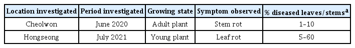
Occurrence of leaf and stem rot on three-leaf ladybell plants grown in fields at two locations of Korea in 2020 and 2021
The leaf rot symptoms started on the petioles of the leaves at the soil line. As the disease progressed, the infected plant parts turned softened and the symptoms advanced upward. Later the entire leaves rotted and blighted (Fig. 1A, B). The stem rot symptoms initially appeared on the stems at or above the soil line. The infected parts of the stems turned brown to dark brown and rotted. As the disease progressed, the infected stems wilted and later blighted (Fig. 1C, D).
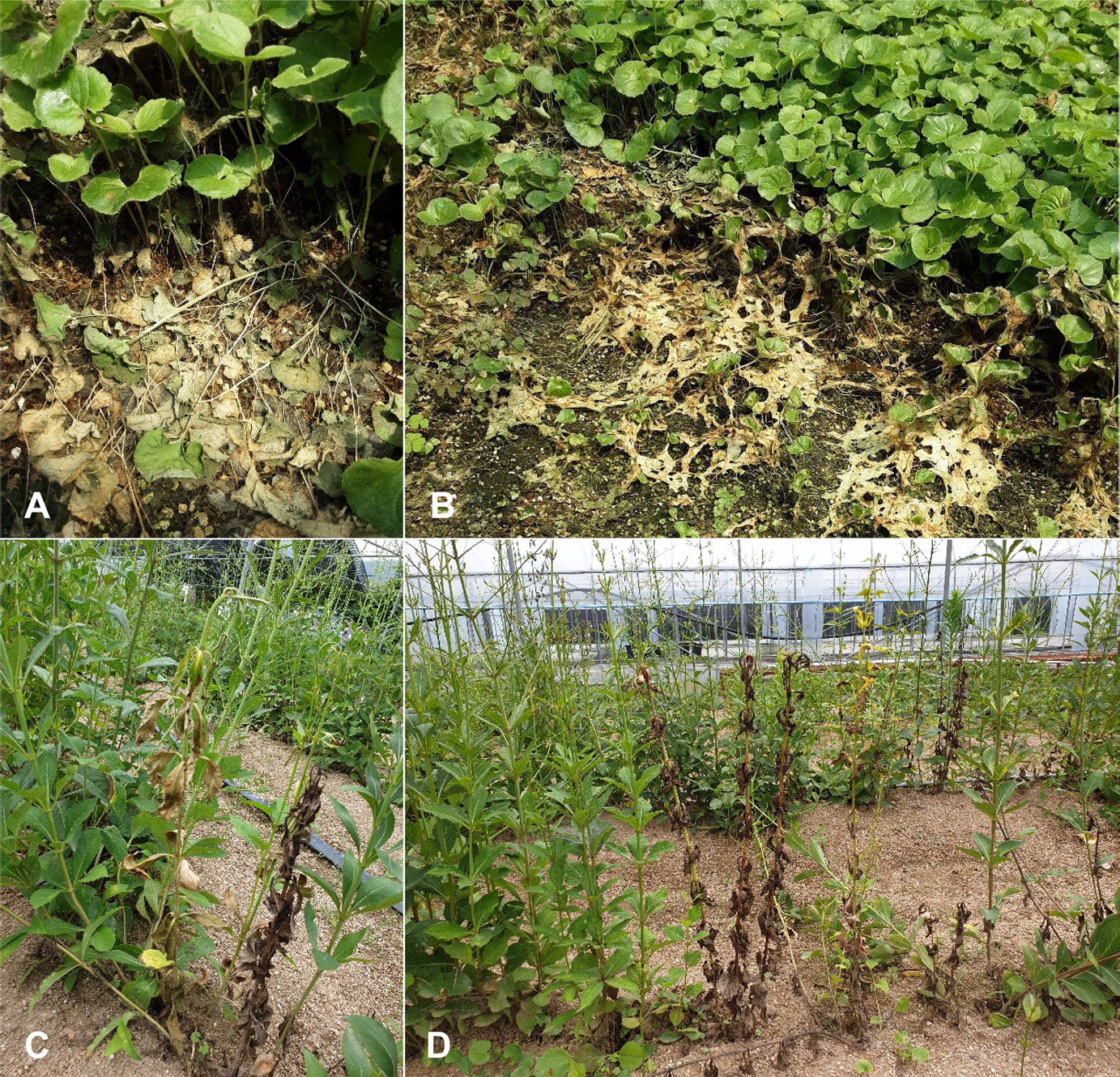
Leaf and stem rot symptoms of three-leaf ladybell plants observed in the fields at two locations of Korea. (A, B) Leaf rot symptoms on young plants grown in Hongseong. (C, D) Stem rot symptoms on adult plants grown in Cheolwon.
Fungal isolates and morphological identification.
Fungi were isolated from diseased leaves and stems of three-leaf ladybell collected during the disease surveys. We obtained 6 fungal isolates each from the leaf rot lesions and the stem rot lesions. Microscopic examination of the isolates showed that the fungal hyphae branched near the distal septa of the hyphal cells, and the hyphae were constricted at the points of the hyphal branches. Hyphal septa were formed at short distance from the points of the hyphal branches. Conidia and clamp connections on the hyphae were not observed. All the examined isolates were identified as R. solani based on their morphological characteristics according to the previous descriptions (Parmeter and Whitney, 1970; Sneh et al., 1991).
Identification of anastomosis groups and cultural types.
Anastomosis tests revealed that the 6 R. solani isolates from the leaf rot lesions of three-leaf ladybell were identified as AG-1, and the other 6 isolates from the stem rot lesions of the plant as AG-2-2. Respective anastomosis reactions between the tested isolates and the tester isolates of R. solani AG-1 and AG-2-2 are shown in Fig. 2A and B. The colony of the R. solani AG-1 isolates cultured on PDA displayed light brown to grayish brown (Fig. 2C). Sclerotia were formed separately or aggregately on the medium. They were grayish brown, subspherical to irregure and measured 0.5‒6.0 mm in diameter. Their surface was mycelioid or wooly. The colony of the R. solani AG-2-2 isolates cultured on PDA displayed brown to dark brown, with concentric zones (Fig. 2D). Sclerotia were absent or rarely formed on the medium, which were dark brown, wooly, and spherical to irregular. The cultural types of the R. solani AG-1 isolates and AG-2-2 isolates were identified as IB and IIIB, respectively, based on the cultural characteristics described in a previous study (Watanabe and Matsuda, 1966).
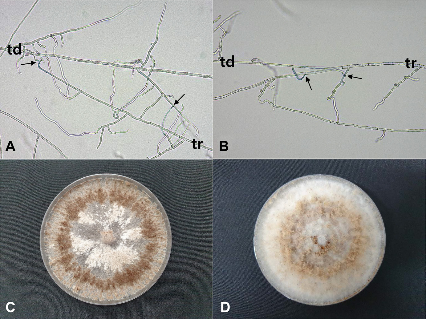
Anastomosis tests of Rhizoctonia solani isolates from three-leaf ladybell plants and cultural appearances of the isolates. (A, B) Anastomosis reactions between the tested isolates (td) and the tester isolates (tr) of R. solani AG-1(IB) and AG-2-2(IIIB), respectively. The arrows in-dicate points of hyphal anastomosis. (C, D) Colonies of R. solani AG-1(IB) and R. solani AG-2-2(IIIB) grown on potato dextrose agar at 25°C for 10 days, respectively.
Result of phylogenetic analyses.
As a result of PCR amplification, 12 isolates of R. solani were amplified to 598‒613 bp. The R. solani isolates were 99‒100% consistent with the two different AG groups by BLASTn analysis. Six isolates from the diseased leaves of three-leaf ladybell belonged to R. solani AG-1(IB), and the other 6 isolates from the diseased stems of the plant to R. solani AG-2-2(IIIB) (Fig. 3). The sequence data of ITS region genes from the 12 isolates of R. solani were deposited in the NCBI GenBank with accession numbers OQ875193‒ OQ875204.
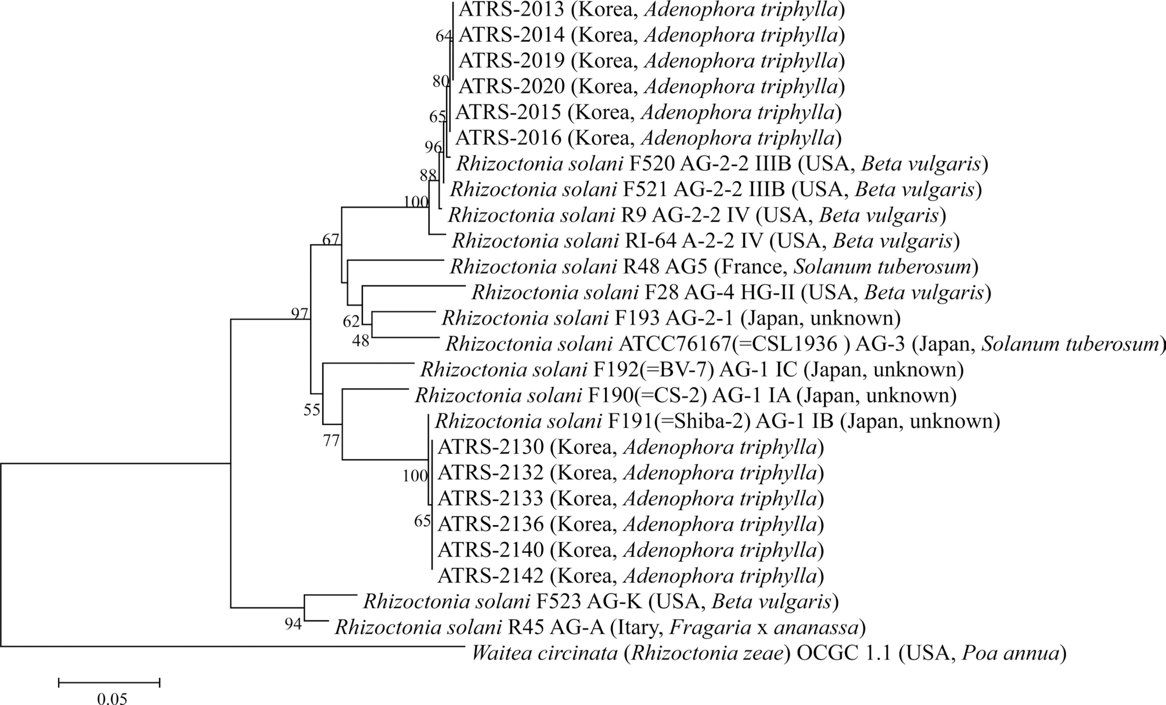
Phylogenetic tree based on the sequences of internal transcribed spacers and intervening 5.8S rDNA region of Rhizoctonia solani isolates from three-leaf ladybell plants and reference species. Sequence data of R. solani strains and Waitea circinata were obtained from the National Center for Biotechnology Information GenBank database. The phylogenetic tree was generated using the neighbor-joining method and Kimura 2-parameter model. Bootstrap support values are given at the nodes. The bar represents the number of nucleotide substitutions per site.
The AG-1(IB) and AG-2-2(IIIB) of the R. solani isolates from three-leaf ladybell plants identified through the rDNA-ITS sequence analysis were consistent with those identified based on anastomosis test and investigation of cultural features of the isolates.
Pathogenicity.
The pathogenicity tests revealed that the isolates of R. solani AG-1(IB) caused only leaf rot symptoms on the inoculated plants of three-leaf ladybell, and those of R. solani AG-2-2(IIIB) leaf rot and stem rot symptoms (Table 2). The symptoms induced by inoculation tests with the two anastomosis groups of R. solani were similar to those observed in the investigated fields (Fig. 4A, B), but no symptoms were induced in the non-inoculated plants (Fig. 4C, D). Reisolation of the inoculated isolates from the lesions was confirmed.
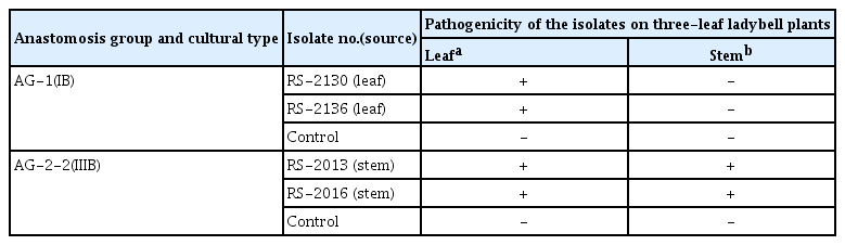
Result of pathogenicity tests of Rhizoctonia solani isolates belonging to two anastomosis groups and cultural types to three-leaf ladybell plants by artificial inoculation
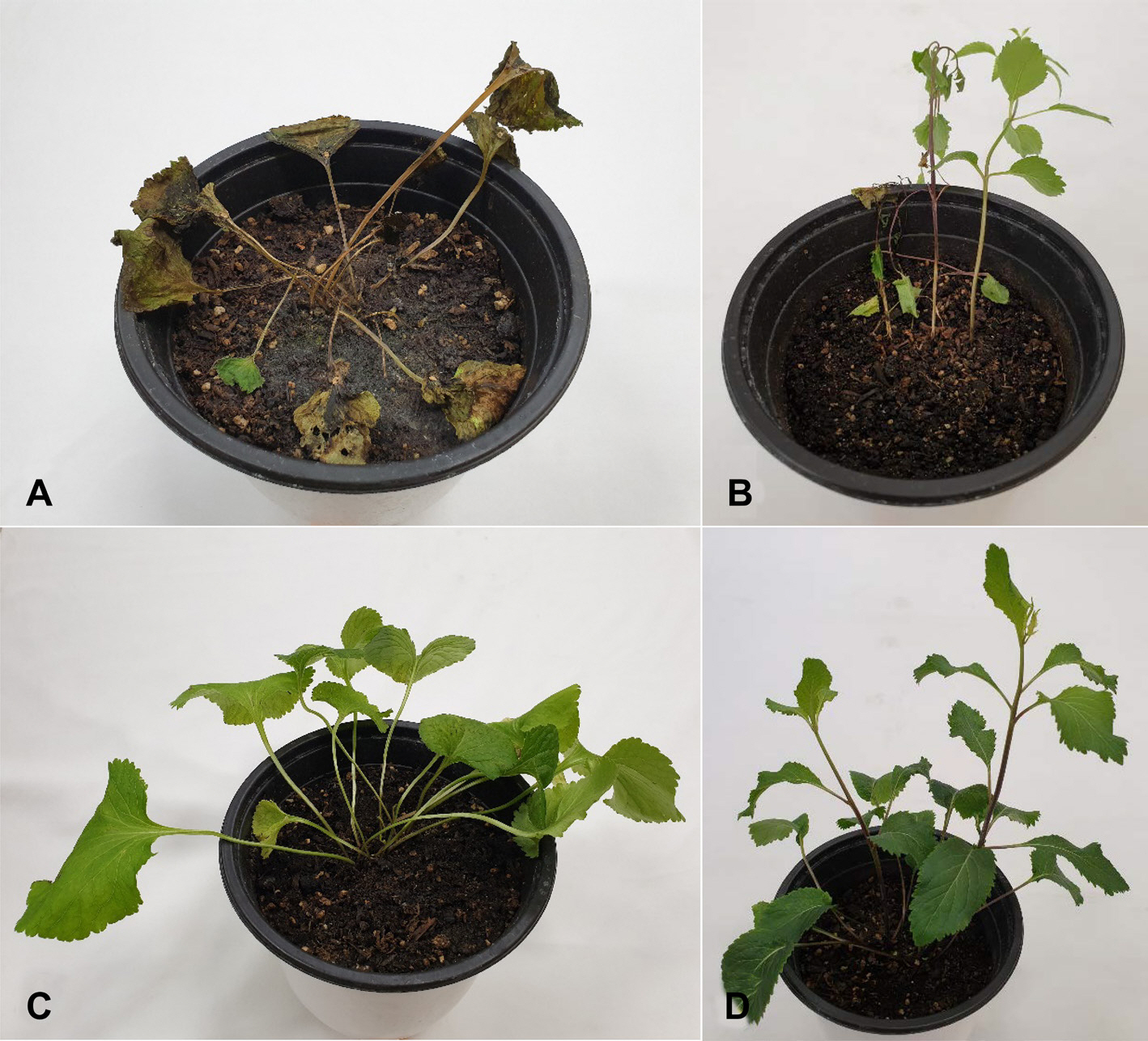
Leaf and stem rot symptoms of three-leaf ladybell plants induced by artificial inoculation tests with two anastomosis groups and cultural types of Rhizoctonia solani isolates. (A) Leaf rot symptoms caused by R. solani AG-1(IB) isolate. (B) Stem rot symptoms caused by R. solani AG-2-2(IIIB) isolate. (C, D) Non-inoculated control plants used in inoculation tests with the isolates of R. solani AG-1(IB) and R. solani AG-2-2(IIIB), respectively.
R. solani causes various diseases in many crops, and anastomosis groups and cultural types of the fungus cause different symptomatic features on the crops (Kim, 1996; Kim et al., 1995). It has been reported that R. solani AG-1(IB) mostly causes damping-off, leaf blight, and leaf rot on the host crops, and R. solani AG-2-2(IIIB) damping-off, root and stem rot, and petiole rot in Korea (Kim, 1996). In this study, R. solani AG-1(IB) was isolated from leaf rot lesions of young three-leaf ladybell plants and induced the leaf rot lesions by inoculation test. In addition, R. solani AG-2-2(IIIB) was isolated from stem rot lesions of adult three-leaf ladybell plants and induced the leaf and stem rot lesions by inoculation test. Therefore, it is considered that R. solani AG-1(IB) could attack only leaves of three-leaf ladybell, but R. solani AG-2-2(IIIB) both leaves and stems of the plant. There has been no report on leaf and stem rot of three-leaf ladybell caused by R. solani. Leaf and stem rot of three-leaf ladybell caused by the two anastomosis groups and cultural types of R. solani is first reported in this study.
Notes
Conflicts of Interest
No potential conflict of interest relevant to this article was reported.
Acknowledgments
This study was supported by a research grant (PJ014507012021) from the Rural Development Administration, Korea.