Res. Plant Dis > Volume 29(1); 2023 > Article
Resazurin 기반 호흡 측정법을 이용한 고추탄저병균에 대한 살균제의 효과 검정

요 약

Resazurin 기반 호흡 측정법으로 strobilurin계 살균제에 대해서 감수성인 고추탄저병균 Colletotrichum acutatum s. lat. 20JDS8에 대한 살균제의 호흡 억제 효과를 조사하였다. Potato dextrose broth에 병원균의 포자를 접종하고 배양하면서 시간별로 상대 형광값(relative fluorescence unit)을 조사한 결과, 12시간 후부터 상승하기 시작하여 24시간 후의 1×10⁴, 1×10⁵, 1×10⁶ spores/ml의 포자 접종구에서 상대 형광값은 1,965.5, 5,412.5, 10,061.0이었다. 병원균을 0, 6, 12, 24시간씩 배양 후에 공시한 살균제를 처리하고, 24시간 후에 상대 형광값을 조사하였다. Dithianon, isopyrazam, pyraclostrobin, fluazinam을 병원균의 포자(0시간), 포자 발아(6시간), 균사 생장(12시간) 단계에 처리할 경우, 각 살균제의 고농도에서 호흡을 90-100% 억제하였다. 하지만 병원균을 24시간 배양한 후에 살균제를 처리할 경우에는 호흡 억제 효과가 크게 감소하였다. Pyraclostrobin 저항성인 C. acutatum s. lat. 20CDJ6에 대해서 pyraclostrobin, azxoystrobin, trifloxystrobin, kresoxim-methyl을 병원균의 모든 생장 단계에 각각 처리하였을 때, 호흡에 대한 억제 효과는 매우 미미하거나, 나타나지 않았다.

ABSTRACT

Resazurin-based microtiter assay was used to evaluate the inhibitory effect of fungicides on the respiration of Colletotrichum acutatum s. lat. 20JDS8 sensitive and 20CDJ6 resistant to strobilurin fungicides. The spores of C. acutatum s. lat. 20JDS8 were inoculated into potato dextrose broth (PDB) at densities of 1×10⁴, 1×10⁵ and 1×10⁶ spores/ml, respectively. The relative fluorescence unit (RFU) of all treatments inoculated at each spore density started to rise after 12 hr of incubation, and were 1,965.5, 5,412.5, and 10,061.0, respectively, after 24 hr of incubation. To evaluate the inhibitory effect of fungicide on the respiration of the pathogen, the spores of the pathogen were inoculated into the PDB and treated with the fungicides 0, 6, 12, and 24 hr after incubation, respectively. After keeping the pathogen culturing for another 24 hr, PrestoBlue reagent was treated into the PDB culturing the pathogen. The RFU of each treatment was examined 1 hr after the reagent was treated. When dithianon, isopyrazam, pyraclostrobin, and fluazinam were treated at high concentrations in the stages of spores (immediately after inoculation [0 hr]), spore germination (after incubation for 6 hr), and hyphal growth (after incubation for 12 hr), the respiration of pathogens was inhibited by 90-100%. When the fungicides were treated after culturing the pathogen for 24 hr, the respiratory inhibitory effects were greatly reduced. With pyraclostrobin-resistant C. acutatum s. lat. 20CDJ6, azxoystrobin, trifloxystrobin and kresoxim-methyl, which have the same mode of action, had very little or no respiratory inhibitory effect in all growth stages of pathogens. Based on the above results, it was thought that the resazurin-based microtiter assay could quickly and accurately evaluate the inhibitory efficacy of the fungicides that inhibited respiration.

서 론

살균제 효과를 검정하는 방법으로는 병원균의 생장 억제 효과를 직접 조사하는 방법과 식물체에서 병 발생을 억제하는 병 방제 효과를 조사하는 방법 등이 있다(Lee 등, 2014; Park과 Kim, 2022; Park 등, 2014). 병원균 생장에 대한 억제 효과를 검정하기 위해서 병원균의 균사생장 또는 포자발아에 대한 억제 효과를 조사하거나, 병원균의 대사 과정에 관여하는 효소 활성에 대한 억제 효과 등을 조사한다. 병원균에 대한 살균제의 효과를 배지에서 검정할 때에는 일반적으로 한천희석법을 사용하여 균사 생장에 대한 억제 효과를 조사하는 데, 많은 시간과 노력, 그리고 병원균을 배양하는 일정한 공간 등이 필요하다. 또한 한천희석법은 병원균에 대한 균사 생장 억제 효과는 떨어지지만 포자 발아 억제 효과가 우수한 살균제의 효과를 정확하게 검정할 수 없는 한계를 가지고 있다. Botrytis cinerea에 대한 kresoxim-methyl, azoxystrobin, folpet 등의 포자 발아 억제 효과를 조사한 EC50값은 0.02, 0.32, 0.15 μ g/ml이지만, 균사 생장에 대한 억제 효과는 EC50값이 0.28, 3.1, 5.5 μ g/ml로 10-30배까지 차이가 났다(Slawecki 등, 2002). 이런 결과는 반대인 경우도 있다. Carbendazim과 pyrimethanil의 포자 발아에 대한 EC50값이 >25.0과 8.6 μ g/ml이었던 반면에, 균사 생장에 대한 EC50값은 0.045와 0.48 μ g/ml이었다. 이처럼 살균제의 효과를 검정하기 위해서 사용하는 방법에 따라 살균제의 효과는 다르게 평가되어질 수 있기 때문에 살균제가 가지는 작용 특성이나 조사하고자 하는 효과에 따라서 적절한 검정 방법을 선택하는 것이 중요하다.
포자 발아 억제 검정법은 세포의 생존에 영향을 미치는 살균제의 효과를 검정할 수 있는 조사 방법 중의 하나이지만, 병원균의 포자를 현미경으로 관찰하여 발아 여부를 결정해야 하기 때문에, 많은 시간과 노력이 소요된다. 따라서, 다수의 균주를 조사하거나, 다수의 화합물을 조사하기에는 적합하지 못하다. 그런데 세포의 생존을 조사하는 방법으로 미토콘드리아의 활성, lactate 탈수소효소나 adenylate kinase의 분비, ATP의 세포 내 함량 등을 측정하는 방법 등이 있다(Miret 등, 2006). 살균제의 효과 검정에 미토콘드리아의 활성을 측정하는 방법을 사용하는데, MTT (3-(4,5-dimethylthiazol-2-yl)-2,5-diphenyltetrazolium bromide)나 resazurin (7-hydroxy-10-oxidophenoxazin-10-ium-3-one) 등과 같은 시약이 사용된다(Boncler 등, 2014; Mosmann, 1983). MTT 검정법은 세포 생존을 조사하기 위해서 확립된 초기의 방법으로, 미토콘드리아의 숙신산(succinic acid) 탈수소효소에 의해서 tetrazolium 염인 MTT가 환원되어 물에 녹지 않는 보라색의 formazan 결정을 만들기 때문에, dimethyl sulfoxide나 HCl/isopropanol 과 같은 용매로 formazan을 추출한 후 정량하여 효과를 검정한다(Mosmann, 1983). 이 과정에서 formazan이 세포에 독성을 나타내기 때문에, 세포 또는 대상 병원균을 배양하면서 연속적으로 생존 여부를 측정할 수 없는 단점이 있다. 또한 MTT 환원 반응은 대사 활동이 활발한 세포에서만 발생하기 때문에, 대사 활동이 활발하지 못한 세포의 경우에는 세포의 수가 동일하게 존재한다고 하더라도 MTT 검정의 결과가 달라질 수 있다(Vega-Avila와 Pugsley, 2011). Resazurin 기반 검정법에서는 처리한 수용성의 resazurin 시약이 환원되며 분홍색을 내는 resorufin을 생성하기 때문에, 환원된 resorufin의 색도와 형광을 측정한다(Lall 등, 2013). 그런데 resazurin은 tetrazolium염과는 다르게 안정적이며 세포에 대하여 독성을 나타내지 않기 때문에, 세포에 처리한 후 계속적인 조사가 가능한 장점을 가지고 있다. Resazurin은 1990년대 후반부터 AlamarBlue라는 상품화된 시약으로 보급되어 사용되기 시작하였다(Vega-Avila 와 Pugsley, 2011). 식물병원균에서도 resazurin 기반의 세포 생존 검정법을 사용하여 살균제의 효과를 검정하였다. 감귤갈색점무늬병균인 Alternaria alternata에 대해서 배지, 포자 밀도, 처리하는 resazurin의 농도 등이 resazurin이 resorufin으로 환원되는 호흡 과정에 미치는 영향을 실험하여, microtiter plate에서 resazurin 기반 호흡 검정법을 확립하였고, quinone outside inhibitor 살균제에 대한 저항성 모니터링을 실시하였다(Vega 등, 2012). 또한 아보카도, 망고, 파파야, 파프리카 등에서 분리한 Colletotrichum gloeosporioidesC. truncatum에 대해서 resazurin 기반 시약인 AlamarBlue를 사용하여 살균제에 대한 반응을 검정하였다(Rampersad와 Teelucksingh, 2012). 또 다른 resazurin 기반 시약 중에 하나인 PrestoBlue는 2011년부터 상품화되어 사용되었는데, 무독성이며 세포막 투과성이 있고, 빛을 쪼이지 않으면 상온에서도 몇 달동안 안정적이라는 장점을 가지고 있다. 특히 아주 적은 세포 수에도 반응할 수 있을 뿐만 아니라, 시약을 처리하고 10분에서 1시간 사이에 반응을 조사할 수 있어서 약제, 화합물, 천연물 등의 세포 독성을 조사하는 실험에 사용되고 있다(Ayyagari 등, 2017; Emter와 Natsch, 2015; Luzak 등, 2022; Oosthuizen 등, 2017). PrestoBlue는 resazurin 기반의 시약으로, 살아있는 세포에 처리하게 되면 미토콘드리아 막 효소들에 의해서 resazurin이 resorufin이라는 물질로 환원하게 된다(Kamiloglu 등, 2020). 이 때 resazurin의 파란색이 새롭게 생성된 resorufin에 의해서 분홍색으로 변하고, 일정 파장에서 resorufin이 형광을 발하기 때문에, 흡광도나 형광을 측정하게 되면 세포의 호흡과 생존을 간접적으로 검정할 수 있다(Boncler 등, 2014).
본 실험에서는 고추에서 분리한 탄저병균을 사용하여 배지와 병원균의 포자 밀도가 resazurin의 환원에 미치는 영향을 조사하였으며, 병원균의 생장 단계별로 살균제를 처리하여 병원균의 호흡에 미치는 살균제의 효과를 검정하였다. 또한 strobilurin계 살균제로서 이미 저항성 발생이 보고된 pyraclostrobin에 대해서 감수성과 저항성인 고추탄저병균을 선발하여(Kim 등, 2019), 작용기작이 다른 살균제의 효과를 검정하였다.

재료 및 방법

실험에 사용한 병원균.

병든 고추에서 단포자 분리한 C. acutatum sensu lato 20JDS8과 C. acutatum s. lat. 20CDJ6을 사용하여 실험하였다. Potato dextrose agar (PDA) 사면 배지에서 배양한 고추탄저병균은 배지 위에 유동 파라핀(White Oil DB-T-1000, MOLKANG, Paju, Korea)을 붓고 실온에서 보관하였다. 실험 중에는 병원균을 PDA 사면 배지에 배양하여 4 o C에서 보관하면서 실험에 사용하였다.

실험에 사용한 살균제.

고추탄저병균에 대한 살균제 효과를 검정하기 위해서 dithianon (a.i. 75%, WP), isopyrazam (a.i. 12.57%, EC), pyraclostrobin (a.i. 20%, WG), fluazinam (a.i. 50%, WP), tebuconazole (a.i. 25%, WP), prochloraz (a.i. 25%, EC) 등 작용기작이 서로 다른 6종의 살균제를 선발하여 실험에 사용하였다. 실험에 사용한 살균제의 농도는 Table 1과 같았다.
Table 1.
Fungicides and their concentrations used in each test method
Fungicide A.I. a (%) and formulation b Test method Concentration (μg/ml) c
Dithianon 75%, WP Agar dilution 100.0, 10.0, 1.0, 0.1
PrestoBlue 100.0, 50.0, 25.0, 12.5, 6.25, 3.13, 1.56, 0.78, 0.39
Isopyrazam 12.57%, SC Agar dilution 10.0, 2.0, 0.4, 0.08, 0.016
PrestoBlue 100.0, 50.0, 25.0, 12.5, 6.25, 3.13, 1.56, 0.78, 0.39
Pyraclostrobin 20%, WG Agar dilution 10.0, 2.0, 0.4, 0.08, 0.016
PrestoBlue 10.0, 5.0, 2.5, 1.25, 0.63, 0.31, 0.16, 0.08, 0.04
Fluazinam 50%, WP Agar dilution 100.0, 10.0, 1.0, 0.1, 0.01
PrestoBlue 100.0, 50.0, 25.0, 12.5, 6.25, 3.13, 1.56, 0.78, 0.39
Tebuconazole 25%, WP Agar dilution 50.0, 5.0, 0.5, 0.05, 0.005
PrestoBlue 50.0, 25.0, 12.5, 6.25, 3.13, 1.56, 0.78, 0.39, 0.20
Prochloraz 25%, EC Agar dilution 4.0, 0.8, 0.16, 0.032, 0.0064
PrestoBlue 4.0, 2.0, 1.0, 0.5, 0.25, 0.13, 0.06, 0.03, 0.02

a A.I. is an abbreviation of active ingredient, which means the active ingredient of the fungicide used.

b The formulations of the fungicides used in this experiment were wettable powder (WP), soluble concentrate (SC), and wettable granule (WG).

c In this experiment, agar dilution method and resazurin-based respiration measurement method were used to evaluate the fungicides, and the concentrations of fungicides treated were shown. In the agar dilution method, the potato dextrose agar as solid medium was poured into a Petri dish, and the fungicide was added at indicated concentration to test the efficacy inhibiting the mycelial growth of Colletotrichum acutatum s. lat. causing pepper anthracnose. In the resazurin-based respiration measurement method, a small amount of liquid medium inoculated with conidia of C. acutatum s. lat. was poured into a microtiter plate, and the fungicide was treated at the concentrations shown. The pathogens were cultured for 24 hr and treated with PrestoBlue reagent to examine the relative fluorescence units.

병원균의 균사생장 억제효과 검정.

실험에 사용한 살균제는 멸균증류수에 현탁하여 살균제의 최종 농도가 Table 1에서와 같은 농도가 되도록 멸균한 배지에 첨가하였다. 세균의 오염을 방지하기 위해 PDA 배지에 300 μ g/ml의 streptomycin을 같이 첨가하고, 직경이 6 cm인 Petri 접시에 부어 살균제 배지를 준비하였다. 고추탄저병균은 25 o C의 PDA 배지에서 7일간 배양한 후, 균총의 선단에서 직경 3 mm의 균사 조각을 떼어 접종원으로 사용하였다. 살균제를 농도별로 첨가한 PDA 배지의 중앙에 균총 선단에서 떼어낸 균사조각의 균사면이 배지를 향하도록 뒤집어서 접종하였다. 병원균을 접종한 배지는 25 o C의 암조건에서 5-7일간 배양하면서 살균제를 첨가하지 않은 배지에서 균총의 직경이 약 40 mm 전후가 되었을 때, 직경을 조사하였고, 아래 식에 의해서 살균제의 균사생장 억제율(%)을 계산하였다.
균사생장 억제율(%)=1-살균제 첨가 배지의 균총의 직경살균제 무첨가 배지의 균총의 직경×100

병원균의 포자 형성.

고추탄저병균을 25 o C의 PDA 배지에 접종하여 암 조건에서 14일간 배양하였다. 배양한 병원균의 공중 균사를 멸균한 슬라이드글라스를 이용하여 긁어 제거한 후, 근자외선을 12시간 간격으로 2일간 조사하여 포자 형성을 유도하였다. 멸균증류수를 배지에 붓고 멸균한 슬라이드글라스로 균총 표면을 긁어 포자를 수확하고, 포자 현탁액을 4겹의 거즈에 여과하여 균사 또는 배지 등의 이물질을 제거하였다. 포자 현탁액을 3,000 rpm에서 15분간 2회 원심분리하여 포자를 세척하고, 현미경 하에서 현탁액의 포자 밀도를 정해진 밀도로 조절하여 사용하였다.

PrestoBlue 시약을 이용한 resorufin의 형광 조사.

고추탄저병균의 포자현탁액을 96 well microtiter plate (96-well cell culture plate, SPL Life Sciences, Pocheon, Korea)의 한 well당 100 μ l씩 분주한 후 25 o C 암 상태에서 배양하였다. 정해진 배양 시간 별로 PrestoBlue 시약을 한 well당 10 μ l씩 처리한 다음, 동일한 조건에서 1시간을 배양하고 각 well의 형광을 조사하였다. 여기 파장(excitation wavelength)과 방출 파장(emission wavelength)을 각각 530 nm와 590 nm로 조정하여 microplate reader (Synergy HTX, Bio Tek, Seoul, Korea)에서 상대 형광값(relative fluorescence unit, RFU)을 측정하였다. 또한 병원균의 균사 생장 정도를 광학현미경(KR-300, Korea Labtech Corporation, Seongnam, Korea)으로 직접 관찰하였다.

배지와 포자 밀도가 resazurin 환원에 미치는 영향.

병원균 배양에는 potato dextrose broth (PDB; Difco, Becton, Dickinson and Company, Sparks, MD, USA)와 Czapek Dox broth (CDB, Difco, Becton, Dickinson and Company), 그리고 각각을 1/5로 희석한 20% PDB와 20% CDB를 사용하였다. 또한 배지에 접종한 고추탄저병균의 최종 포자 밀도는 1×10⁴, 1×10⁵, 1×10⁶ spores/ml로 조절하여 실험하였다. Resorufin의 형광은 병원균을 배양하면서 정해진 시간별로 조사하였다.

PrestoBlue 검정법을 통한 살균제의 효과 검정.

고추탄저병균의 포자현탁액(포자밀도; 1×10⁵ spores/ml)을 96 well microtiter plate의 well당 99 μ l씩 분주한 후 25 o C의 암 상태 배양기에서 0, 6, 12, 24시간 배양하고, 살균제를 각각 1 μ l씩 처리하였다. 처리한 살균제의 최종 농도는 Table 1과 같았다. 병원균 배양 0시간 후 살균제 처리는 병원균을 96-well microtiter plate에 접종함과 동시에 살균제를 처리하였다. 무처리 대조구는 살균제 대신 멸균증류수를 처리하였다. 살균제를 처리한 고추탄저병균은 살균제를 처리한 직후부터 25 o C 암 상태에서 24시간을 배양하고 PrestoBlue 시약(Invitrogen, Life Technologies Corporation, OR, USA)을 well당 10 μ l씩 처리한 다음, 동일한 조건에서 1시간을 배양하고 RFU를 조사하였고, 동시에 광학현미경을 사용하여 균사의 생장 정도를 조사하였다. 형광값을 이용한 살균제의 억제효과는 아래식을 이용하여 계산하였다.
살균제의 억제효과(%)=(1PTt24PtPt24Pt)×100
P t: t시간 후 포자 현탁액의 형광값
P t→24: t시간 배양 후 살균제를 처리하지 않고 다시 24시간 배양 후 측정한 형광값
PT t→24: t시간 배양 후 살균제를 처리하고 다시 24시간 배양 후 측정한 형광값

Pyraclostrobin 저항성 고추탄저병균에 대한 살균제의 억제 효과 및 교차 저항성 검정.

Pyraclostrobin 감수성 C. acutatum s. lat. 20JDS8과 저항성인 20CDJ6에 대해서 Table 1에서와 같은 농도로 dithianon, isopyrazam, fluazinam, tebuconazole 을 처리하고, 병원균에 대한 억제효과를 PrestoBlue 검정법으로 조사하였다. 또한 azoxystrobin (a.i. 21.7%, SC), kresoxim-methyl (a.i. 50%, WG), trifloxystrobin (a.i. 22%, SC)을 선발하여 pyraclostrobin과의 교차 저항성 여부를 조사하였다.

결과 및 고찰

배지와 포자 밀도가 resazurin의 환원에 미치는 영향.

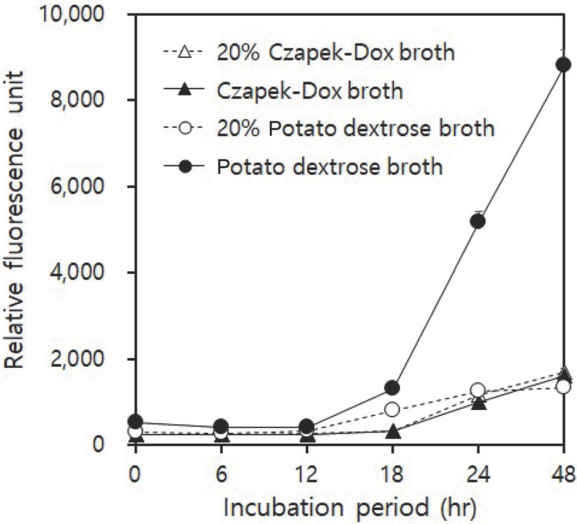
실험에 사용한 PDB와 20% PDB, 그리고 CDB와 20% CDB 중에서 고추탄저병균을 PDB에서 배양하였을 때, RFU가 가장 높게 상승하였다. Fig. 1에서 보는 것과 같이 병원균의 포자 밀도를 1×10⁵ spores/ml로 접종한 PDB에서 12시간 이후부터 형광이 증가하기 시작하여 18시간 이후에 급격하게 증가하였다. 배양 24시간 후에는 PDB 배지에서 RFU가 다른 배지보다 4배 이상 상승하였다.
Fig. 1.
The influence of incubation media and period on relative fluorescence units of PrestoBlue reagent used to monitor the growth of Colletotrichum acutatum s. lat. 20JDS8 causing pepper anthracnose. Data were collected from experiments with 3 replication and ex-pressed as mean±standard error. The readings of relative fluorescence unit were made in fluorescence units at 530/590 nm.
RPD-2023-29-1-11f1.jpg
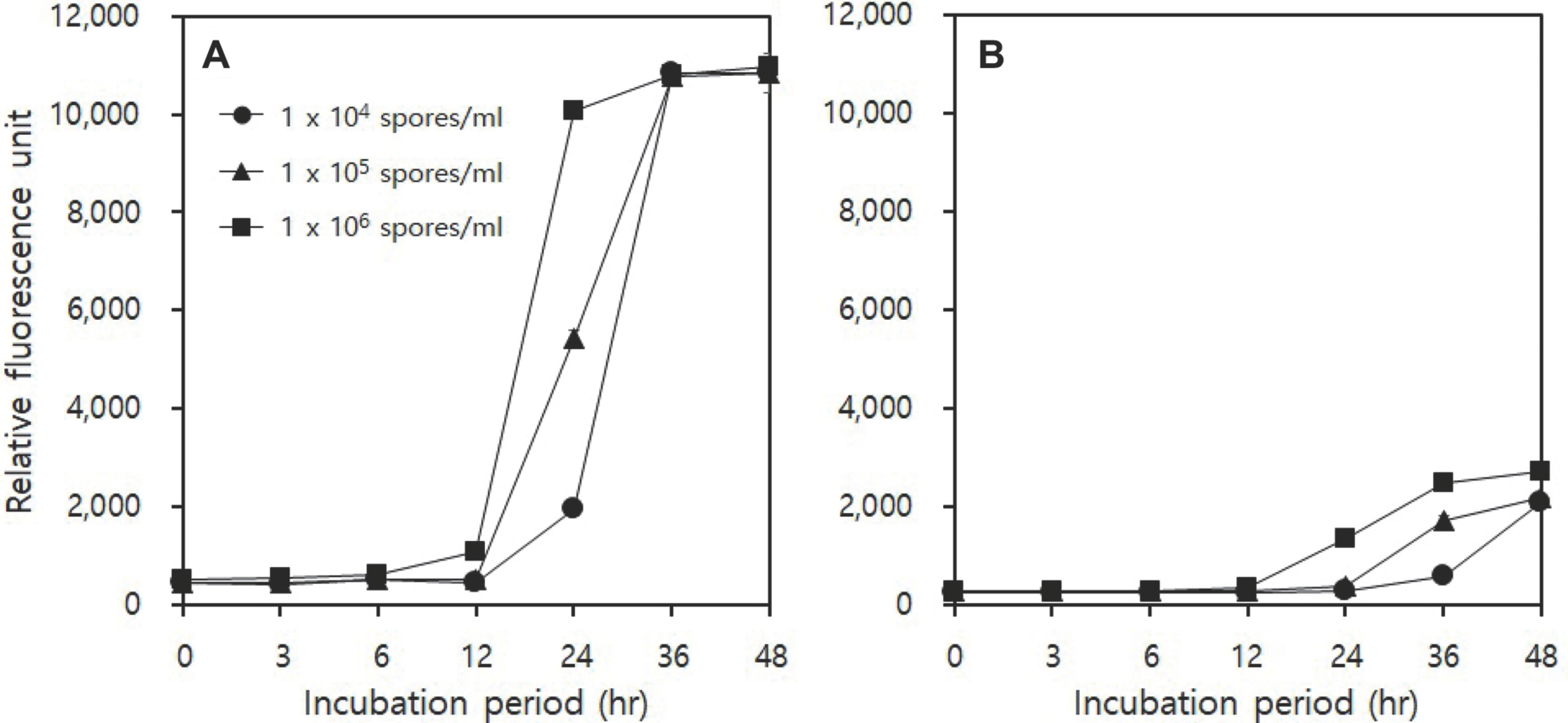
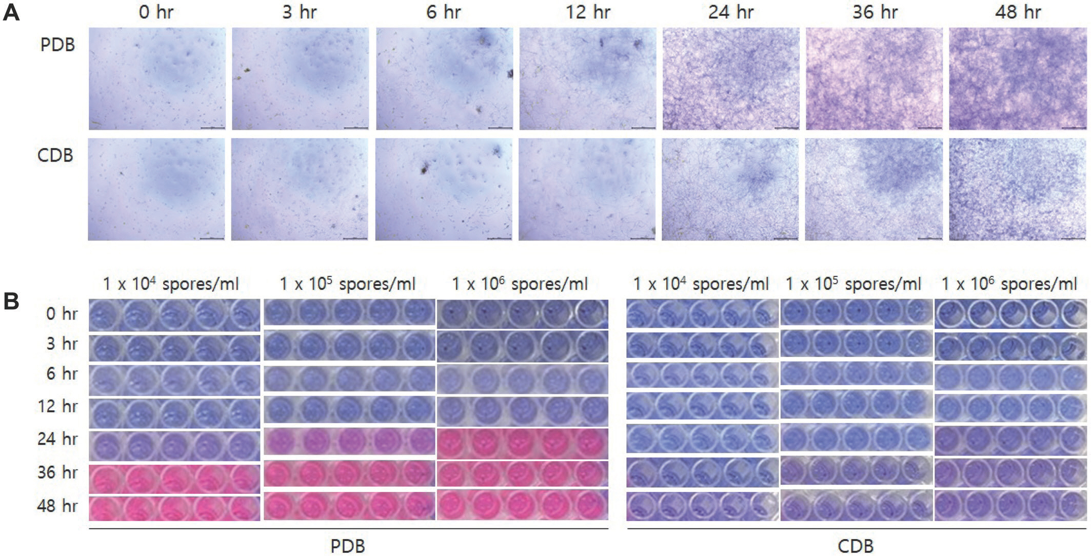
PDB와 CDB 배지에 병원균의 포자 밀도를 다르게 하여 접종하였을 때에도, 배양 12시간 후부터 PDB와 CDB의 RFU 에 현저한 차이가 나타나기 시작하였으며, PDB의 경우 24시간 후에는 접종한 병원균의 밀도에 따라서 뚜렷한 차이가 나기 시작하였다(Fig. 2). PDB에 병원균의 포자 밀도를 1×10⁴spores/ml로 접종하고 24시간 배양한 처리구의 RFU는 1,956이었으며, 1×10⁵ spores/ml와 1×10⁶ spores/ml로 접종한 처리구는 5,412와 10,061로 나타났다. 하지만 CDB 배지에서는 접종한 모든 포자 밀도에서 RFU는 낮게 나타났는데, 1×10⁶ spores/ml의 처리구에서 조차 24시간과 48시간 후의 RFU 는 1,372과 2,707이었다. 병원균을 1×10⁶ spores/ml로 접종한 microtiter plate에서 병원균의 모습을 현미경으로 관찰하면, Fig. 3A에서 보는 것과 같이 병원균을 12시간 배양한 후부터 CDB보다 PDB에서 포자 발아와 균사 생장이 빠르게 진행됨을 관찰할 수 있었다. 병원균의 포자가 발아하고 균사의 생장이 활발해지는 12시간 후부터 처리한 PrestoBlue 시약에서 resazurin이 resorufin으로 환원되면서 배지의 색깔이 파란색에서 붉은색으로 변화되는 모습을 육안으로 확인할 수 있었으며, 24시간 배양한 이후부터 배지의 색깔 변화가 뚜렷하게 나타나기 시작하였다(Fig. 3B).
Fig. 2.
Effects of media and spore densities on relative fluorescence unit of Colletotrichum acutatum s. lat. 20JDS8 causing pepper anthracnose by lapse of incubation time. In this experiment, as media were used potato dextrose broth (A) and Czapek Dox broth (B).
RPD-2023-29-1-11f2.jpg
Fig. 3.
The growth of Colletotrichum acutatum s. lat. 20JDS8 (A) observed over time after inoculating the pathogen into the media as potato dextrose broth (PDB) and Czapek Dox broth (CDB), and the color change of media (B) treated with PrestoBlue reagent according to incubation time. The spore density of the pathogen inoculated into PDB and CDB, where spore germination and mycelial growth of pathogens were observed under a microscope, was 1×10⁶ spores/ml.
RPD-2023-29-1-11f3.jpg
Resazurin이 환원하여 나타나는 resorufin의 형광을 측정한 RFU는 접종한 병원균 포자 밀도에 따라서 달랐으며, 이런 RFU 의 차이는 검정하고자 하는 살균제의 효과에 영향을 미친다(EspinelIngroff와 Kerkering, 1991; Vega 등, 2012). 배지 또한 배양하는 병원균이 생장하는데 필요한 양분을 충분히 함유하고 있어야 할 뿐만 아니라, 에너지를 만들어 내는 과정에서 대체 호흡이나 혐기성 발효 등이 발생하지 않아야 한다(Spiegel 과 Stammler, 2006). 경우에 따라서 배지는 검정하는 화합물의 수용해도나 안정성에도 영향을 미친다(Pijls 등, 1994). 따라서 살균제의 효과 검정을 위해서는 검정하는 방법과 병원균 등에 따라서 표준화 작업이 필요하다. Vega 등(2012)은 Alternaria alternata에 대한 호흡 저해 살균제의 효과를 검정하기 위해서 resazurin의 환원 억제 정도를 조사하였는데, 완전 배지에서 접종하는 포자 농도에 따른 resazurin의 환원 억제 정도가 가장 큰 폭으로 나타났다. PDB의 yeast peptone dextrose broth에서 환원 억제 정도가 완전배지보다 크게 나타났지만, 포자 밀도에 따른 억제 폭이 크지 않았기 때문에, 완전배지를 사용하였다. 하지만 본 실험에서는 Figs. 12에서 보는 것과 같이 PDB에서 C. acutatum s. lat.의 RFU가 높게 나타났을 뿐만 아니라 포자 밀도와 배양 시간 등에 따라서 큰 폭의 차이가 나타났기 때문에 PDB가 호흡 저해 살균제의 효과를 검정하는 데 적합할 것으로 판단하였다.

병원균의 생육 단계별로 처리한 살균제의 호흡 억제 효과.

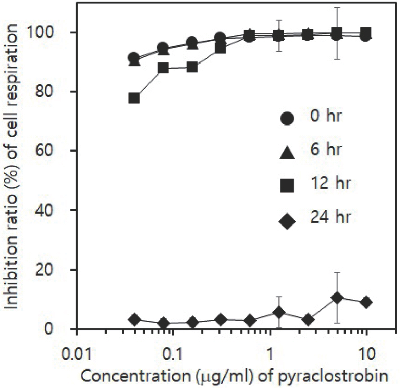
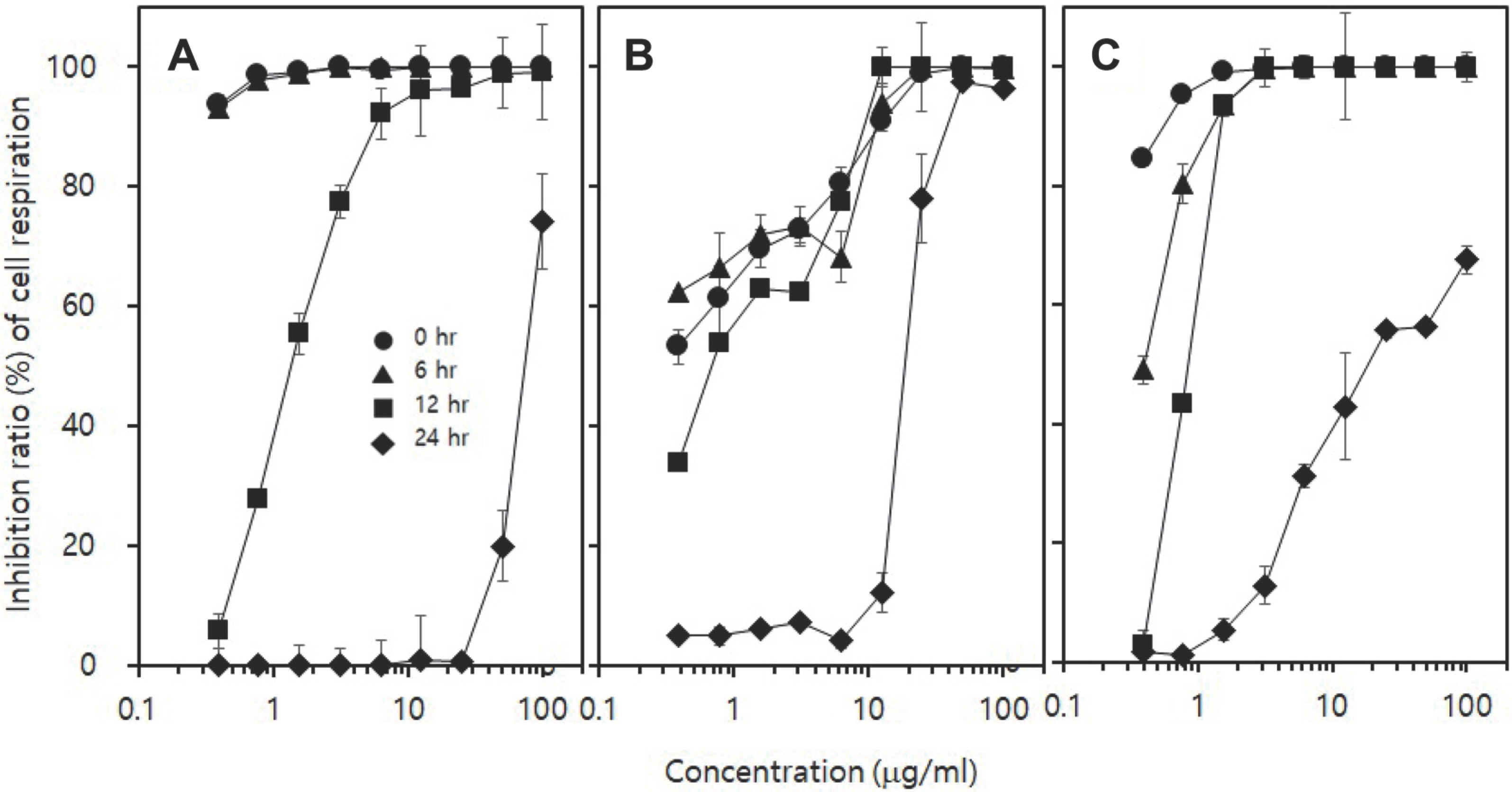
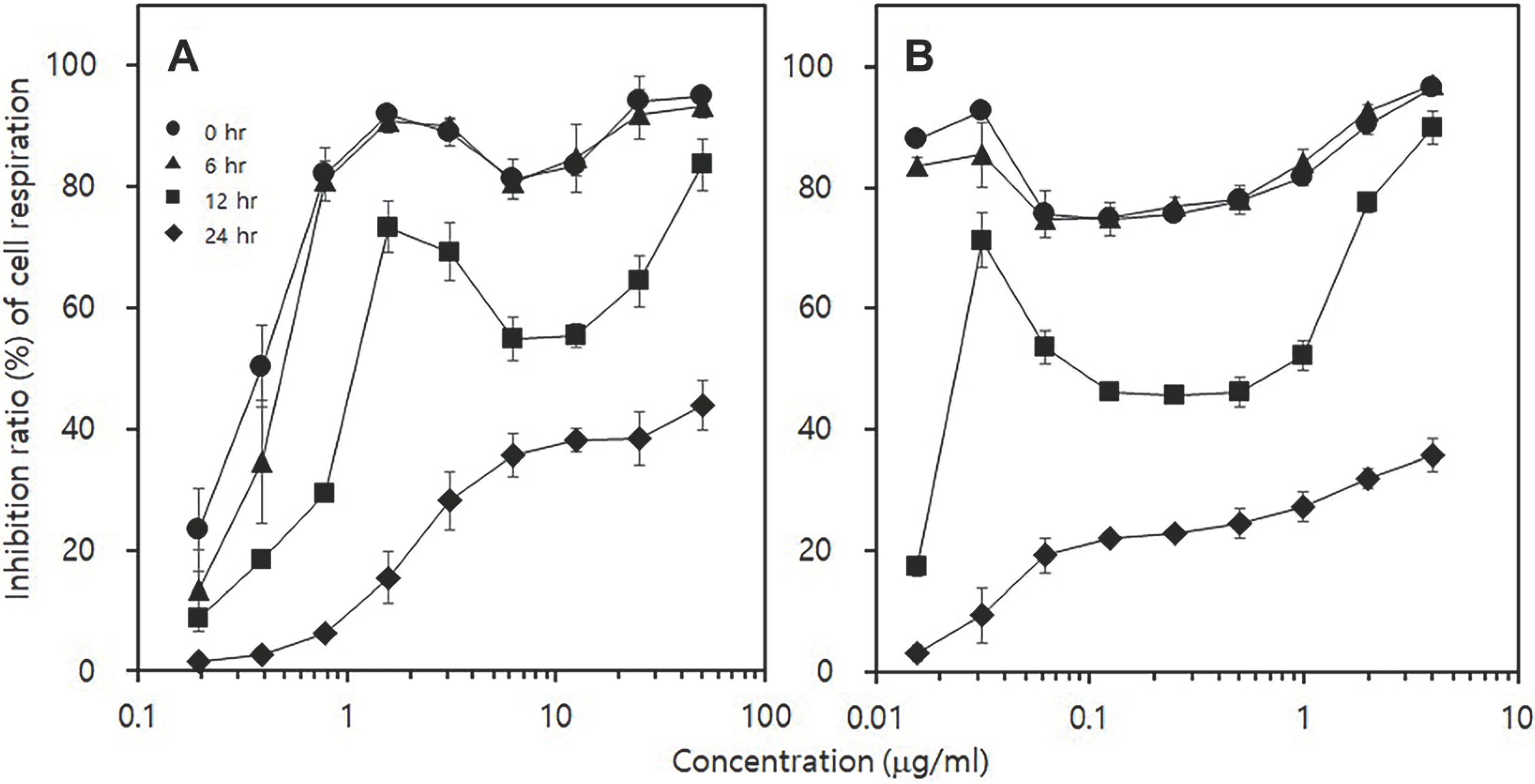
고추탄저병균의 생육 단계를 포자 단계, 포자 발아 단계, 균사 생장 단계, 균사체 형성 단계로 나누어 pyraclostrobin 을 처리하였다(Fig. 4). Fig. 3A에서 포자 단계는 20JDS8의 포자를 PDB에 접종한 직후를 나타내며, 포자 발아 단계는 병원균의 포자를 PDB에 접종하고 6시간 배양하여 70% 이상의 포자가 발아한 단계를 말하였다. 균사 생장 단계는 PDB에 접종한 포자를 12시간 배양하여 발아관이 균사로 생장하는 단계를, 그리고 균사체 형성 단계는 24시간 배양하여 96-well micrititer plate의 well에 균사체가 가득 생성된 단계를 나타내었다. Pyraclostrobin을 병원균의 각 생장 단계에 처리하였을 때, 병원균에 대한 호흡 저해 효과는 포자 단계부터 균사 생장 단계에 처리하였을 때에 나타났다. 하지만 균사체 형성 단계에 pyraclostrobin을 처리하였을 때에는 병원균에 대한 호흡 저해 효과가 전혀 나타나지 않았다. 보호 살균제(카 군)인 dithianon과 세포의 호흡을 저해하는 다2 군과 다5 군에 속하는 isopyrazam과 fluazinam 역시 균사 생장 단계인 포자를 접종하고 12시간 배양한 후에 처리하였을 때까지는 농도에 따라서 차이는 있었지만, 병원균의 호흡을 효과적으로 억제하였다. 하지만 24시간 배양한 후인 균사체 형성 단계에는 10 μ g/ml 이하의 처리 농도에서는 효과가 매우 미미하였다(Fig. 5). 병원균의 세포막 구성 성분인 ergosterol 생합성을 저해하는 살균제(사1 군)인 tebuconazole과 prochloraz는 포자 단계에 처리하였을 때에도 호흡을 100% 억제하지 못하였다(Fig. 6). Dithianon의 경우 포자 단계에 0.039 μ g/ml를 처리하여도 90% 이상의 호흡 억제 효과를 보였던 반면에, 균사 생장 억제 효과를 조사한 한천희석법의 결과를 보면 100 μ g/ml에서 20JDS8의 균사 생장을 60.5%밖에 억제하지 못하였다(Fig. 7D).
Fig. 4.
Inhibitory efficacy of pyraclostrobin against Colletotrichum acutatum s. lat. 20JDS8 causing pepper anthracnose according to the application time. Pyraclostrobin was treated with each indicated concentration at each growth stage of the pathogen such as spore stage, germinating stage of spore, hyphal growth stage and constructing stage of mycelia. The spore stage was the time when the spores were inoculated into the potato dextrose broth (PDB), and the germinating stage of spores was the time when the spores inoculated in the PDB were cultured for 6 hr and more than 70% of the spores germinated. The hyphal growth stage was the period in which germ tubes grew into long hyphae after culturing for 12 hr, and the constructing stage of mycelia was the period when the wells of the microtiter plate were filled with mycelia. Respiration of the pathogen was measured after being treated with a fungicide and then incubated for another 24 hr. Relative fluorescence unit was measured by adjusting the excitation and emission wavelengths to 530 nm and 590 nm, 1 hr after treatment with PrestoBlue reagent.
RPD-2023-29-1-11f4.jpg
Fig. 5.
Inhibitory efficacy of dithianon (A), isopyrazam (B), and fluazinam (C) against Colletotrichum acutatum s. lat. 20JDS8 causing pepper anthracnose according to the application time. Fungicides were treated with each indicated concentration at each growth stage such as spore stage, germinating stage of spore, hyphal growth stage and constructing stage of mycelia. Respiration of the pathogen was measured after being treated with a fungicide and then incubated for another 24 hr. One hour after treatment with PrestoBlue reagent, relative fluorescence unit was measured by adjusting the excitation and emission wavelengths to 530 nm and 590 nm.
RPD-2023-29-1-11f5.jpg
Fig. 6.
Inhibitory efficacy of tebuconazole (A) and prochloraz (B) against Colletotrichum acutatum s. lat. 20JDS8 causing pepper anthracnose according to the application time. Fungicides were treated with each indicated concentration at each growth stage such as spore stage, germinating stage of spore, hyphal growth stage and constructing stage of mycelia. Respiration of the pathogen was measured after being treated with a fungicide and then incubated for another 24 hr. One hour after treatment with PrestoBlue reagent, relative fluorescence unit was measured by adjusting the excitation and emission wavelengths to 530 nm and 590 nm.
RPD-2023-29-1-11f6.jpg
Fig. 7.
Inhibitory effect of fungicides on mycelial growth of Colletotrichum acutatum s. lat. 20JDS8 sensitive to pyraclostrobin and 20CDJ6 resistant to pyraclostrobin on potato dextrose agar amended with each fungicide. Inhibition ratio (%) of mycelial growth was evaluated by using an agar dilution method. (A) Pyraclostrobin. (B) Isopyrazam. (C) Fluazinam. (D) Dithianon. (E) Tebuconazole. (F) Prochloraz.
RPD-2023-29-1-11f7.jpg
이처럼 특정한 작용 특성을 지닌 살균제는 검정법에 따라서 병원균에 대한 효과에 차이가 크다. Dithianon과 같이 보호 살균제인 보르도액은 187 μ g/ml의 처리에서 고추탄저병균의 포자 발아를 100% 억제하였지만, 동일한 농도에서 병원균의 균사 생장은 전혀 억제하지 못하였으며, 포자 발아를 100% 억제하는 187 μ g/ml의 농도보다 25배나 높은 4,685 μ g/ml의 농도를 처리해야 균사의 생장을 100% 억제할 수 있었다(Park 등, 2014). 그런데 tebuconazole과 prochloraz처럼 균사 생장 억제 효과가 더 우수한 살균제의 경우에는 dithianon과는 반대로 PrestoBlue 시약을 사용하는 검정법보다는 한천희석법을 통하여 균사 생장 억제 효과를 조사하는 것이 더 우수한 효과를 얻을 수 있다. 따라서 살균제의 효과를 검정하고자 할 때는 검정하고자 하는 살균제의 작용 기작과 특성 등을 고려하여 적합한 검정법을 선택하는 것이 살균제의 효과를 정확하게 측정하는 데 중요하다.

Pyraclostrobin 감수성균과 저항성균의 호흡 저해 효과.

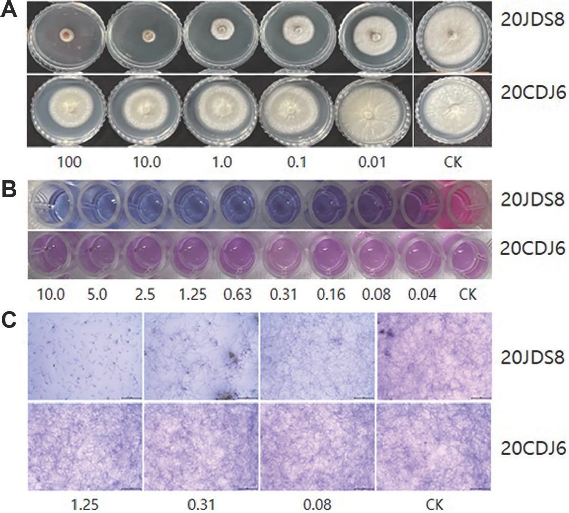
Pyraclostrobin 저항성인 C. acutatum s. lat. 20CDJ6과 감수성인 20JDS8에 대한 균사생장 효과를 한천희석법으로 실험한 결과, Fig. 8A에서 보는 것과 같이 처리한 pyraclostrobin의 농도가 높아짐에 따라 20JDS8의 균사생장은 억제되었지만, 20CDJ6의 균사생장은 거의 억제되지 않았다. 이러한 차이는 PrestoBlue 시약을 사용하여 병원균의 호흡을 측정하여도 동일한 경향으로 나타났다(Fig. 8B). 처음에 파란색이었던 배지의 색이 감수성균인 20JDS8의 살균제 무처리구에서는 분홍색으로 변하였으나, pyraclostrobin의 처리구에서는 파란색 그대로 남아있었다. 이는 처리한 PrestoBlue 시약의 resazurin이 미토콘드리아의 환원 효소들에 의해서 resorufin으로 환원되며 붉은색으로 변화되어야 하는데, pyraclostrobin에 의해서 효소의 활성이 억제되며 환원되지 못한 결과로 생각된다. 그러나 저항성균인 20CDJ6에서는 10 μ g/ml의 pyraclostrobin을 처리하였는데 불구하고 배지의 파란색이 분홍색으로 변화하는 것을 보면, 처리한 살균제가 resazurin을 환원시키는 효소의 활성을 저해하지 못한 것을 알 수 있었다. 현미경으로 병원균의 생장을 조사한 결과에서도 병원균의 생장에 큰 차이가 있었다(Fig. 8C). Pyraclostrobin 을 1.25 μg/ml로 처리한 경우, 20JDS8은 병원균이 발아한 직후 더 이상 생장하지 못하였지만, 20CDJ6의 생장은 억제되지 않았다. Fig. 8에서 보는 것과 같이 pyraclostrobin 저항성 균주인 20CDJ6은 한천희석법, PrestoBlue 시약을 이용한 호흡 측정법과 현미경 검정법에서 감수성 균주인 20JDS8과 뚜렷한 차이를 보였다. 세 가지의 검정법 중에서 한천희석법은 결과를 얻기 위해서 약 5일 정도가 소요되기 때문에, 호흡 측정법이나 현미경 관찰법과 비교하여 결과를 얻기까지 긴 시간이 필요하다. 뿐만 아니라, 실험하는 Perti 접시를 보관하고 배양해야 하는 공간도 필요하다. 현미경 관찰법은 호흡 측정법과 비교하여, 검정하는데 많은 시간과 노력이 필요하다. 따라서 PrestoBlue 시약을 이용한 호흡 측정법은 호흡 억제 살균제의 효과를 빠르고 쉽게 검정할 수 있을 뿐만 아니라, 포장에서 분리한 균주의 저항성 검정에도 효과적으로 사용할 할 수 있을 것으로 생각한다.
Fig. 8.
Response of Colletotrichum acutatum causing pepper anthracnose to pyraclostrobin according to the assay method. In this experiment, C. acutatum 20JDS8 sensitive to pyraclostrobin and 20CDJ6 resistant to the fungicide, were used. Numbers in each assay represent the concentrations (μg/ml) of pyraclostrobin. (A) Agar dilution method investigating the mycelial growth inhibitory effect of pyraclostrobin. (B) Respiration assay method using PrestoBlue reagent. (C) Microscopic observation for investigating the change of pathogen morphology. In the agar dilution method, pathogens were inoculated on potato dextrose agar medium amended with fungicide at indicated concentrations, and cultured at 25°C for 5 days. Then the diameter of the mycelial colonies were measured. In the respiration assay method, the pathogens were treated with pyraclostrobin by indicated concentrations at that time when the pathogens were inoculated into potato dextrose broth. After culturing the pathogen for 24 hr, the PrestoBlue reagent was treated for 1 hr and fluorescence was measured. In light microscopy, it was observed after treatment with pyraclostrobin and incubation for 24 hr.
RPD-2023-29-1-11f8.jpg
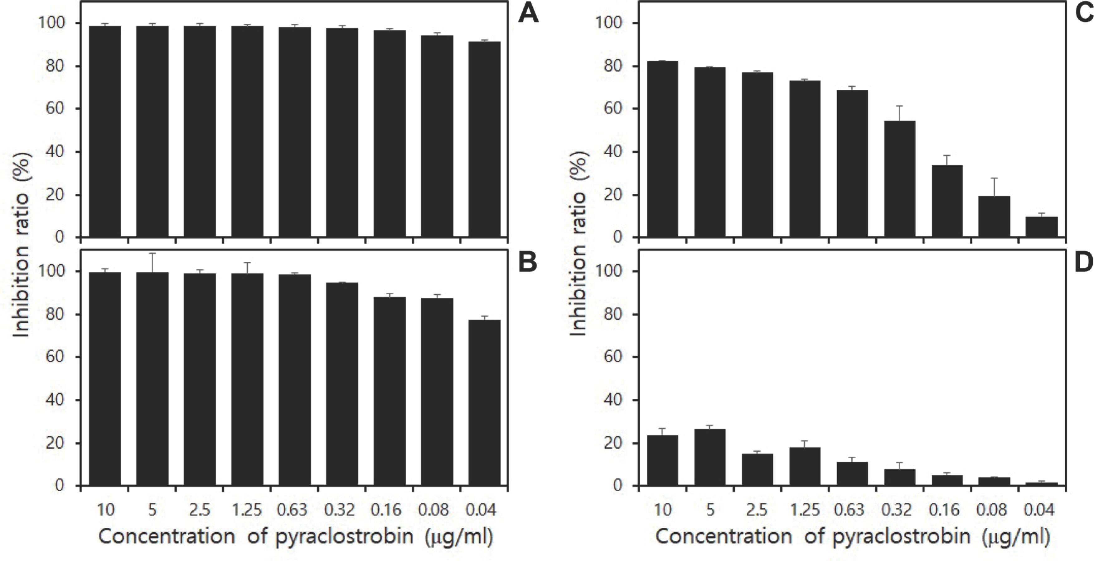
C. acutatum s. lat. 20CDJ6과 20JDS8의 생육 단계에 따라 pyraclostrobin을 처리한 후, 살균제의 효과를 호흡 측정법으로 조사하면 Fig. 9와 같이 뚜렷한 차이가 나타났다. 병원균의 포자를 접종함과 동시에 살균제를 처리하였을 때, 감수성 균주인 20JDS8에 대한 10.0과 0.04 μ g/ml의 억제 효과는 98.5%와 91.1%이었지만, 저항성인 20CDJ6에 대해서는 82.3%와 9.8%의 효과를 보였다(Fig. 9A, C). 병원균을 12시간 배양하고 살균제를 처리하였을 때, 20JDS8에 대한 억제효과는 99.7%와 77.7%이었지만, 20CDJ6에 대해서는 23.8%와 1.6%의 효과를 보였을 뿐이었다(Fig. 9B, D). 20JDS8과 20CDJ6 균주를 모두 PDB 배지에서 24시간 배양한 균총 형성 단계에 pyraclostrobin을 처리한 경우, 감수성과 저항성 균주 모두에서 호흡을 억제하는 효과가 미미하였다.
Fig. 9.
Respiratory inhibition effect of Colletotrichum acutatum s. lat. 20JDS8 (A, B) 20CDJ6 (C, D) to pyraclostrobin using PrestoBlue assay. C. acutatum s. lat. 20JDS8 was sensitive to pyraclostrobin, and 20CDJ6 resistant the fungicide. Pyraclostrobin was treated at indicated concentrations at the same time as the spores of the pathogen were inoculated into the potato dextrose broth (A, C) and after 12 hr of incubation after the spores were inoculated (B, D). The pathogen was cultured for 24 hr after treatment with the fungicide. For measuring relative fluorescence unit (RFU), PrestoBlue reagent was treated after 24 hr of incubation, and RFU was measured 1 hr after treatment with the reagent. The respiratory inhibition effect was calculated by comparing the RFU treated with the fungicide to that of the untreated control.
RPD-2023-29-1-11f9.jpg
Strobilurin계 저항성 병원균과 같이 특정 유전자의 점 돌연변이에 의해서 발생하는 살균제 저항성은 유전자 염기서열을 이용하는 allele-specific polymerase chain reaction (PCR), PCR-based restriction fragment length polymorphism (PCR-RFLP) 등과 같은 분자생물학적인 검정법으로 신속하고 정확하게 검정할 수 있다(Ma와 Michailides, 2005). Cytochrome b 유전자의 143번째 아미노산인 alanine (GTG)이 glycine (GAG)으로 치환되는 점 돌연변이에 의해서 발생하는 pyraclostrobin 저항성 고추탄저병균도 allele-specific PCR이나 PCR-RFLP와 같은 분자생물학적 검정법으로 검정이 가능하다(Isa와 Kim, 2022; Kim 등, 2019). 하지만 병원균에 따라서는 129번째의 phenylalanine이 leucine으로 치환되어 저항성이 발생하기도 하고, 저항성균의 발생 기작이 점 돌연변이와는 관계가 없는 경우도 있다(Hsieh 등, 2013; Kim 등, 2003; Pasche 등, 2005). 저항성 기작이 143번째 아미노산의 점 돌연변이가 아닌 다른 위치의 점 돌연변이이나 다른 기작에 의해서 발생한 저항성균은 cyt b 유전자의 G143A 돌연변이를 기반으로 제작된 primer에 의해서는 저항성균의 유전자를 검정할 수 없다. 따라서 각 저항성균의 기작을 기반으로 하는 분자생물학적인 검정법이 다시 확립되어야 한다. 하지만 PrestoBlue 시약을 이용한 호흡 측정법은 저항성 기작과 관계없이 strobilurin계 살균제에 대한 저항성균을 모두 검정할 수 있는 장점을 가지고 있다(Vega 등, 2012).

Strobilurin계 살균제 간의 교차 저항성.

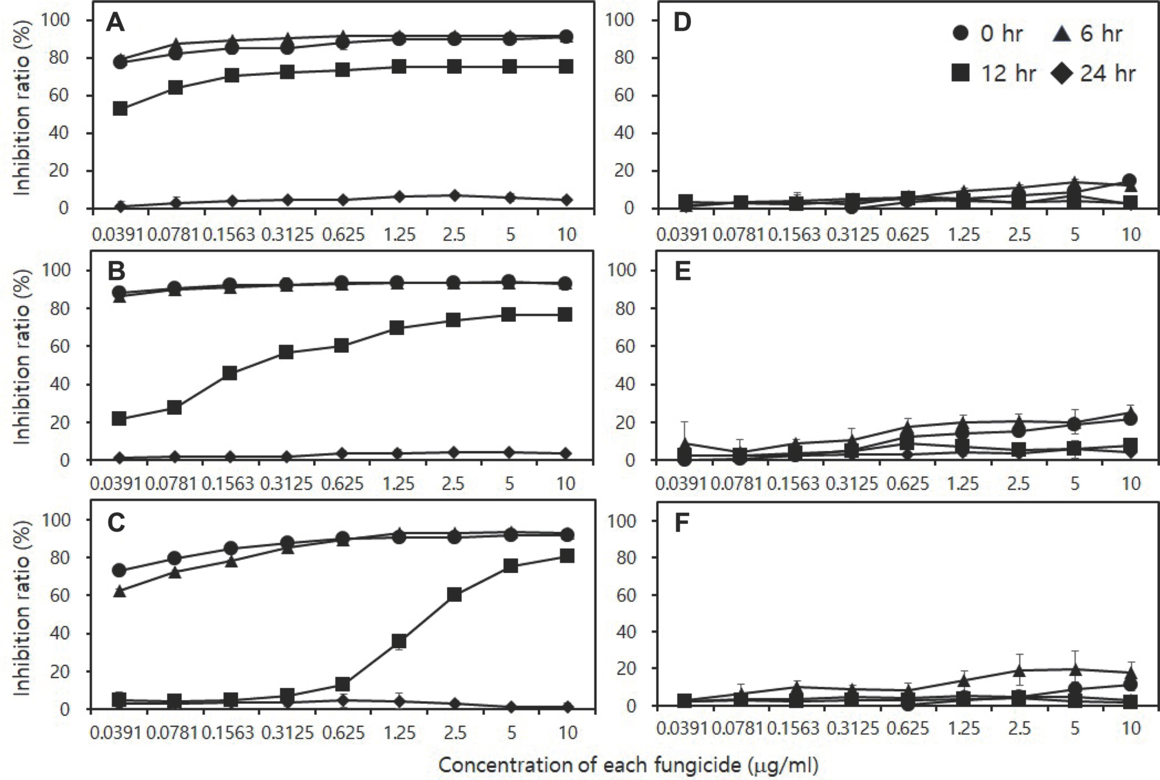
Pyraclostrobin 과 동일한 작용기작을 갖는 azoxystrobin, trifloxystrobin, kresoxim-methyl의 호흡 저해 효과를 확인한 결과, pyraclostrobin 감수성 20JDS8에 대한 살균제의 효과는 확인되었지만, 저항성 20CDJ6에 대한 효과는 병원균을 접종하면서 살균제를 동시에 처리하는 포자 단계 처리에서조차도 미미하였다(Fig. 10). Azoxystrobin의 경우 20JDS8에 대해서 병원균을 접종하면서 또는 6시간 배양한 후에 살균제를 처리하였을 때, 가장 낮은 처리농도이었던 0.04 μ g/ml에서도 호흡에 대한 억제효과가 79.4%를 보였는데, 12시간 배양한 후에 처리한 경우에는 억제효과가 53.2%로 감소하였고, 24시간 배양 후에 처리한 경우에는 호흡에 대한 억제효과가 거의 나타나지 않았다. 배양 시간별로 처리한 trifloxystrobin과 kresoxim-methyl의 20JDS8 균주에 대한 호흡 억제 양상은 azoxystrobin과 유사하였다. 하지만 각 살균제 간에 병원균에 대한 억제 효과에는 차이가 있었는데, 20JDS8을 12시간 배양하고 각 살균제를 0.63 μ g/ml로 처리하였을 때, azoxystrobin, trifloxystrobin, kresoxim-methyl의 억제효과는 73.8%, 12.8%, 60.1%로 살균제에 따라서 효과에 차이가 있었다. 실험에 사용한 3종의 살균제 모두 병원균을 24시간 배양한 후에 처리할 경우에는 병원균의 호흡을 전혀 억제하지 못하였다. 저항성 균주인 20CDJ6에 대해서는 3종의 살균제 모두 병원균을 접종함과 동시에 처리하였을 때에도 병원균 호흡에 대한 억제 효과가 매우 미미하였다.
Fig. 10.
Inhibitory effect (%) of strobilurin fungicides included into quinone outside inhibitors on the cell respiration of Colletotrichum acutatum 20JDS8 (A- C) and 20CDJ6 (D- F). Fungicides were used in this experiment as azoxystrobin (A, D), trifloxysytrobin (B, E), and kresoxim-methyl (C, F). Isolates of the pepper pathogen used in this experiment were C. acutatum s. lat. 20JDS8, which is sensitive to pyraclostrobin, and C. acutatum s. lat. 20CDJ6, which is resistant.
RPD-2023-29-1-11f10.jpg
병원균의 세포 호흡을 저해하는 작용기작을 가진 살균제나 작용점이 다양한 보호 살균제의 효과를 한천희석법을 사용하여 검정할 경우, 병원균에 대한 살균제의 병 방제 효과보다 낮은 효과가 나타날 수 있다. 하지만 PrestoBlue 시약을 이용한 호흡 측정법을 사용하여 효과를 검정할 경우, 본 실험의 결과에서 보는 것처럼 균사 생장 억제 효과가 낮은 살균제의 경우에 높은 호흡 억제 효과를 얻을 수 있었다. 이런 호흡 억제 효과는 살균제가 갖는 병 방제 효과와도 높은 상관 관계를 보이기 때문에, 호흡 저해 살균제나 보호 살균제의 in vitro 활성 검정에는 PrestoBlue 시약을 사용하는 호흡 측정법이 효율적일 것으로 생각한다. 또한 이 방법은 소량의 살균제나 화합물을 가지고서도 검정이 가능하기 때문에, 시료가 소량인 천연물질이나 신규로 합성한 화합물의 검정에도 유용할 것으로 생각한다.

NOTES

Conflicts of Interest

No potential conflicted interest relevant to this article was reported.

Acknowledgments

This work was supported by the Korean Institute of Planning and Evaluation for Technology in Food, Agriculture, Forestry (IPET) through the Crop Viruses and Pests Response Industry Technology Development Program, funded by the Ministry of Agriculture, Food, and Rural Affairs (MAFRA; Grant No. 320042-5).

REFERENCES

Ayyagari, V. N., Hsieh, T.-H. J., Diaz-Sylvester, P. L. and Brard, L. 2017. Evaluation of the cytotoxicity of the Bithionol - cisplatin combination in a panel of human ovarian cancer cell lines. BMC Cancer 17: 49.
crossref pmid pmc pdf
Boncler, M., Różalski, M., Krajewska, U., Podsędek, A. and Watala, C. 2014. Comparison of PrestoBlue and MTT assays of cellular viability in the assessment of anti-proliferative effects of plant extracts on human endothelial cells. J. Pharmacol. Toxicol. Methods 69: 9-16.
crossref pmid
Emter, R. and Natsch, A. 2015. A fast Resazurin-based live viability assay is equivalent to the MTT-test in the KeratinoSens assay. Toxicol. In Vitro 29: 688-693.
crossref pmid
EspinelIngroff, A. and Kerkering, T. M. 1991. Spectrophotometric method of inoculum preparation for the in vitro susceptibility testing of filamentous fungi. J. Clin. Microbiol. 29: 393-394.
crossref pmid pmc pdf
Hsieh, C.-H., Chung, W.-C., Chen, Y.-N. and Chung, W.-H. 2013. Phylogenetic diversity and sensitivity to MBI and QoI fungicides of Magnaporthe oryzae in Taiwan. J. Pestic. Sci. 38: 194-199.
crossref
Isa, D. A. and Kim, H. T. 2022. Cytochrome b gene-based assay for monitoring the resistance of Colletotrichum spp. to pyraclostrobin. Plant Pathol. J. 38: 616-628.
crossref pmid pmc pdf
Kamiloglu, S., Sari, G., Ozdal, T. and Capanoglu, E. 2020. Guidelines for cell viability assays. Food Front. 1: 332-349.
crossref pdf
Kim, S., Min, J. and Kim, H. T. 2019. Occurrence and mechanism of fungicide resistance in Colletotrichum acutatum causing pepper anthracnose against pyraclostrobin. Korean J. Pestic. Sci. 23: 202-211.
crossref
Kim, Y.-S., Dixon, E. W., Vincelli, P. and Farman, M. L. 2003. Field resistance to strobilurin (QoI) fungicides in Pyricularia grisea caused by mutations in the mitochondrial cytochrome b gene. Phyto-pathology 93: 891-900.
crossref
Lall, N., Henley-Simith, C. J., De Canha, M. N., Oosthuizen, C. B. and Berrington, D. 2013. Viability reagent, PrestoBlue, in comparison with other available reagents, utilized in cytotoxicity and anti-microbial assays. Int. J. Microbiol. 2013: 420601.
crossref pmid pmc pdf
Lee, S. M., Jang, H. S. and Kim, H. T. 2014. In vitro fruit assay for the evaluation of fungicide activity against pepper anthracnose. Korean J. Pestic. Sci. 18: 115-121. (In Korean)
crossref
Luzak, B., Siarkiewicz, P. and Boncler, M. 2022. An evaluation of a new high-sensitivity PrestoBlue assay for measuring cell viability and drug cytotoxicity using EA.hy926 endothelial cells. Toxicol. In Vitro 83: 105407.
crossref pmid
Ma, Z. and Michailides, T. J. 2005. Advances in understanding molecular mechanisms of fungicide resistance and molecular detection of resistant genotypes in phytopathogenic fungi. Crop Prot. 24: 853-863.
crossref
Miret, S., De Groene, E. M. and Klaffke, W. 2006. Comparison of in vitro assays of cellular toxicity in the human hepatic cell line HepG2. J. Biomol. Screen. 11: 184-193.
crossref pmid pdf
Mosmann, T. 1983. Rapid colorimetric assay for cellular growth and survival: application to proliferation and cytotoxicity assays. J. Immunol. Methods 65: 55-63.
crossref pmid
Oosthuizen, C., Arbach, M., Meyer, D., Hamilton, C. and Lall, N. 2017. Diallyl polysulfides from Allium sativum as immunomodulators, hepatoprotectors, and antimycobacterial agents. J. Med. Food 20: 685-690.
crossref pmid
Park, S. and Kim, H. T. 2022. Cross-resistance of Colletotrichum acutatum s. lat. to strobilurin fungicides and inhibitory effect of fungicides with other mechanisms on C. acutatum s. lat. resistant to pyraclostrobin. Res. Plant Dis. 28: 122-131. (In Korean)
crossref pdf
Park, S.-J., Lee, S.-M., Gwon, H.-W., Lee, H. and Kim, H. T. 2014. Control efficacy of Bordeaux mixture against pepper anthracnose. Korean J. Pestic. Sci. 18: 168-174. (In Korean)
crossref
Pasche, J. S., Piche, L. M. and Gudmestad, N. C. 2005. Effect of the F129L mutation in Alternaria solani on fungicides affecting mitochondrial respiration. Plant Dis. 89: 269-278.
crossref pmid
Pijls, C. F. N., Shaw, M. W. and Parker, A. 1994. A rapid test to evaluate in vitro sensitivity of Septoria tritici to flutriafol, using a microtitre plate reader. Plant Pathol. 43: 726-732.
crossref
Rampersad, S. N. and Teelucksingh, L. D. 2012. Differential responses of Colletotrichum gloeosporioides and C. truncatum isolates from different hosts to multiple fungicides based on two assays. Plant Dis. 96: 1526-1536.
crossref pmid
Slawecki, R. A., Ryan, E. P. and Young, D. H. 2022. Novel fungitoxicity assays for inhibition of germination-associated adhesion of Botrytis cinerea and Puccinia recondita spores. Appl. Environ. Microbiol. 68: 597-601.
crossref pmid pmc pdf
Spiegel, J. and Stammler, G. 2006. Baseline sensitivity of Monilinia laxa and M. fructigena to pyraclostrobin and boscalid. J. Plant Dis. Prot. 113: 199-206.
crossref pdf
Vega, B., Liberti, D., Harmon, P. F. and Dewdney, M. M. 2012. A rapid resazurin-based microtiter assay to evaluate QoI sensitivity for Alternaria alternata isolates and their molecular characterization. Plant Dis. 96: 1262-1270.
crossref pmid
Vega-Avila, E. and Pugsley, M. K. 2011. An overview of colorimetric assay methods used to assess survival or proliferation of mammalian cells. Proc. West. Pharmacol. Soc. 54: 10-14.
pmid


ABOUT
BROWSE ARTICLES
EDITORIAL POLICY
FOR CONTRIBUTORS
Editorial Office
Rm,904 (New Bldg.) The Korean Science & Technology Center 22, Teheran-ro 7-gil, Gangnam-gu, Seoul 06130, Korea
Tel: +82-2-557-9360    Fax: +82-2-557-9361    E-mail: paper@kspp.org                

Copyright © 2024 by The Korean Society of Plant Pathology.

Developed in M2PI

Close layer
prev next