A Reliable Reverse Transcription Loop-Mediated Isothermal Amplification Assay for Detecting Apple stem grooving virus in Pear
Article information
Trans Abstract
Apple stem grooving virus (ASGV) is a high-risk viral pathogen that infects many types of fruit trees, especially pear and apple, and causes serious economic losses across the globe. Thus, rapid and reliable detection assay is needed to identify ASGV infection and prevent its spread. A reliable reverse transcription loop-mediated isothermal amplification (RT-LAMP) was developed, optimize, and evaluated for the coding region of coat protein of ASGV in pear leaf. The developed RT-LAMP facilitated the simple screening of ASGV using visible fluorescence and electrophoresis. The optimized reaction conditions for the RT-LAMP were 63°C for 50 min, and the results showed high specificity and 100-fold greater sensitivity than the reverse transcription polymerase chain reaction. In addition, the reliability of the RT-LAMP was validated using field-collected pear leaves. Furthermore, the potential application of paper-based RNA isolation, combined with RT-LAMP, was also evaluated for detecting ASGV from field-collected samples. These assays could be widely applied to ASGV detection in field conditions and to virus-free certification programs.
Introduction
Apple stem grooving virus (ASGV) is a species of genus Capillovirus in family Betaflexviridae and has a wide range of hosts, including apple, pear, cherry, citrus, lily, soybean, and tomato (Bhardwaj and Hallan, 2019; Canales et al., 2021). Furthermore, ASGV has been reported in both single and co-infections in fruit trees. Even though ASGV infections can remain latent, the virus often induces severe symptoms (e.g., necrotic spot vein yellow, interveinal mottling, leaf deformation, stem grooving, tree decline, and the deformation on graft unions) in sensitive cultivars. ASGV is commonly spread through the use of infected propagation materials, and no natural insect vectors have been reported to date. Thus, the virus is a high-risk pathogen that requires the establishment of a certification program in Korea for virus detection and control, as well as for the maintenance and distribution of virus-free germplasm and plantlets. However, the establishment of such a program is critically dependent on the ability to detect ASGV in crop plants and germplasm.
Currently, the detection of ASGV in apple, pear, and other fruit trees typically involves immunodiagnostic methods or reverse transcription polymerase chain reaction (RT-PCR) (Ji et al., 2013; Yao et al., 2014). However, these serological and molecular diagnostic methods are limited by relatively long reaction times (2−3 hr) and requirements for specialized molecular biology equipment (e.g., thermal cycler) and skilled researchers. Thus, a simpler detection method that also exhibits high specificity, sensitivity, and reliability is urgently needed. Although a recombinase polymerase amplification method was recently reported as a more rapid (~40 min) detection method for ASGV in pear, the method was also reported to frequently yield non-specific amplification bands, as a result of low reaction temperatures (37−42°C) (Kim et al., 2018).
The application of loop-mediated isothermal amplification (LAMP) have facilitated the early detection of a variety of viral pathogens, including apple stem pitting virus (ASPV), apple chlorotic leaf spot virus (ACLSV), and peach latent mosaic viroid (PLMVd), in fruit trees (Lee and Jeong, 2020; Lu et al., 2018; Peng et al., 2017). Although LAMP requires the development of multiple (four to six) primers that can recognize target sequences in multiplexed reactions, the resulting assays are relatively simple (constant temperature: 60−65°C; no specialized equipment), specific, sensitive, and rapid (30−60 min). In addition, LAMP products can be visualized using color dyes (e.g., SYBR Green and hydroxyl naphthol blue) (Notomi et al., 2000).
The present study was performed to develop, optimize, and evaluate a reverse transcription (RT)-LAMP assay for detecting ASGV in pear trees in Korea. In addition, the use of a paper-based RNA extraction method for facilitating on-site LAMP detection was evaluated.
Materials and Methods
Plant samples. In June–October of both 2017 and 2018, fresh ASGV-infected pear leaf samples were collected from orchards in Namyangju, Naju, Sangju, Ulsan, and Cheonan, with most leaves being collected from ‘Niitaka’ pear trees (Kim et al., 2019).
Isolation of total RNA. Total RNA was extracted from pear samples following to Kim et al. (2019) method and stored at –20°C. For the paper-based extraction of RNA, 50 mg pear leaf samples were each ground and extracted using a tissue grinder pestle and 250 μl extraction buffer for 1 min. A disc of Whatman No. 1 filter paper was then incubated at each extract solution for ≥5 sec and transferred to 400 μl wash buffer for 1 min (Zou et al., 2017), after which the paper discs containing RNA were transferred to RT-LAMP reaction mixtures.
RT-PCR. ASGV was detected using RT-PCR with diagnostic primers and RNA extracted from pear leaf samples (Kim et al., 2019). ACLSV, ASPV, and nad5 were also RT-PCR-amplified using previously reported primer sets (Kim et al., 2019).
Design of ASGV-specific primers for RT-LAMP assay. Twenty ASGV coat protein sequences were obtained from NCBI GenBank, and sequences were aligned using ClustalW in BioEdit v. 7.0.5.3. Four RT-LAMP oligonucleotide primers (Table 1), corresponding to the conserved ASGV coat protein gene regions, were then generated using PrimersExplorer V5 software, with default settings (accession no. LC475148) (Supplementary Fig. 1).
Optimization of RT-LAMP. One-step RT-LAMP was conducted with reaction mixtures that each contained 2 μl of total RNA template, 0.4 μM ASGV F3/B3, 1.6 μM ASGV FIP/BIP, 10× LAMP buffer, 6 μl of 10 mM dNTPs (iNtRON, Seongnam, Korea), 4.5 μl of 5 M betaine, 1 μl of Bst DNA polymerase (8 U/μl, Enzynomics, Daejeon, Korea), 0.2 μl of AMV RTase (600u; Promega, Madison, WI, USA), 1 μl recombinant RNase inhibitor (5,000 U; Takara, Tokyo, Japan), and diethylpyrocarbonate-treated distilled water up to a total of 25 volumes. Optimal temperature condition was evaluated by incubating RT-LAMP reaction mixtures in a water bath at 54°C, 57°C, 60°C, 63°C, 66°C, 69°C, 72°C, and 75°C for 60 min, followed by at 80°C for 5 min in a water bath to terminate the reactions. Meanwhile, the optimal reaction time was evaluated by incubating RT-LAMP reaction mixtures at an optimal temperature of 63°C for 30, 40, 50, and 60 min, followed by at 80°C for 5 min.
In all cases, the RT-LAMP results were tested the presence of ASGV by direct visual observation after the incubation of 100× SYBR Green I (Invitrogen, Carlsbad, CA, USA). Green coloration indicated that reactions were positive for ASGV RNA, whereas red coloration indicated that reactions were negative for ASGV RNA. The RT-LAMP results (5 μl each) were also analyzed by 2% (w/v) agarose gel electrophoresis.
RT-LAMP specificity and sensitivity. The specificities of the RT-LAMP and RT-PCR for ASGV detection were evaluated using RNA extracted from pear leaf samples infected with either ACLSV or ASPV, which are also major pathogens of pear in Korea, and from pear leaf samples free of all three viruses (negative control), with three replicate RT-LAMP and RT-PCR reactions per sample. Meanwhile, the sensitivities of the methods for ASGV detection were evaluated using a 10-fold serial dilution (1.5×10⁻¹ to 1.5×10⁻⁸ μg) of total RNA from ASGV-infected samples and total RNA from healthy pear leaf samples as negative controls, with three replicate reactions per sample.
Results
Optimization of the RT-LAMP assay. The total RNA samples were used to assess the optimal RT-LAMP incubation temperature and time for ASGV detection. To determine optimal reaction time for ASGV detection, RT-LAMP was performed at 54°C, 57°C, 60°C, 63°C, 66°C, 69°C, 72°C, and 75°C for 50 min. Intermediate reaction temperatures (60–69°C) yielded relatively high amplicon concentrations (typical ladder-like patterns and visual color changes), both lower (54°C and 57°C) and higher (72°C and 75°C) temperatures yielded little to no amplification (Fig. 1). With results of amplicon shape and intensity, 63°C was identified as the optimal reaction temperature. To determine optimal reaction time for ASGV detection, RT-LAMP was conducted at 63°C for 1, 5, 10, 20, 30, 40, 50, and 60 min. Longer reaction times (40–60 min) yielded relatively high amplicon concentrations, whereas shorter reaction times failed to yield any amplification (Fig. 2). Based on amplicon intensity and preference for shorter reaction time, 50 min was identified as the optimal reaction time. Therefore, all subsequent RT-LAMP assays were performed under optimal conditions at 63°C for 50 min.
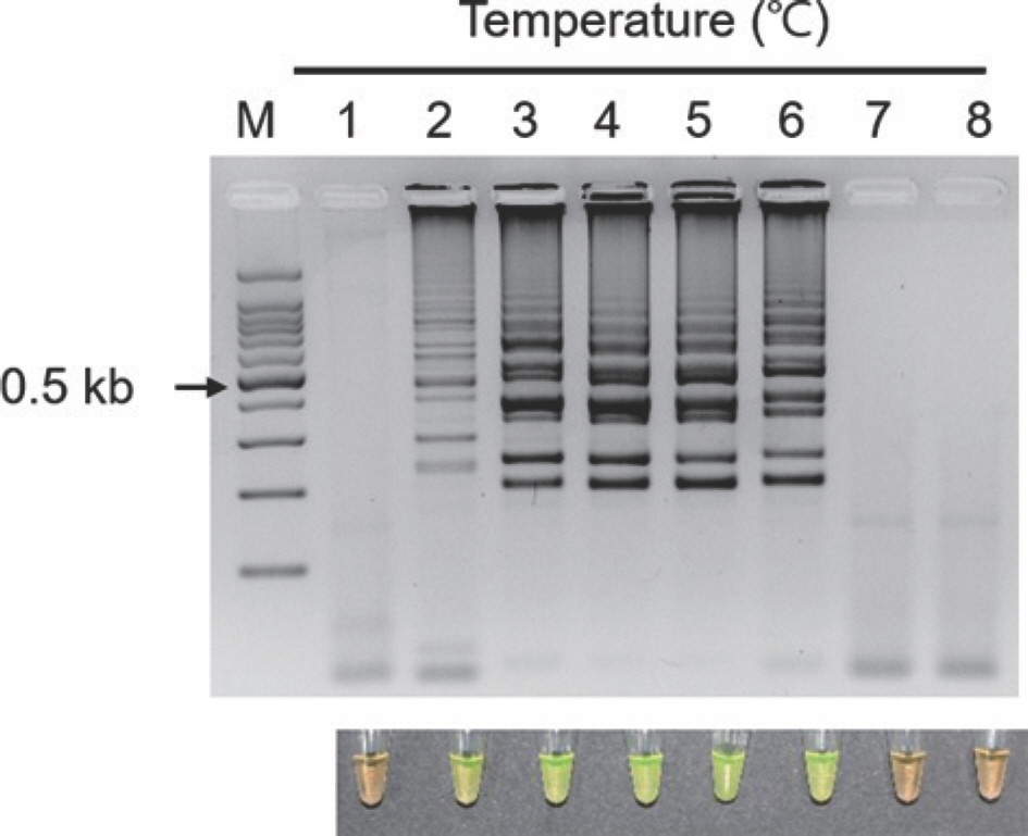
Effect of reverse transcription loop-mediated isothermal amplification (RT-LAMP) incubation temperature on the detection of Apple stem grooving virus (ASGV). RT-LAMP results were evaluated by 2% agarose electrophoresis and SYBR Green I. Orange color represents negative and yellow color represents positive. Lane M, 100-bp DNA ladder; lanes 1–8: 54°C, 57°C, 60°C, 63°C, 66°C, 69°C, 72°C, and 75°C, respectively.
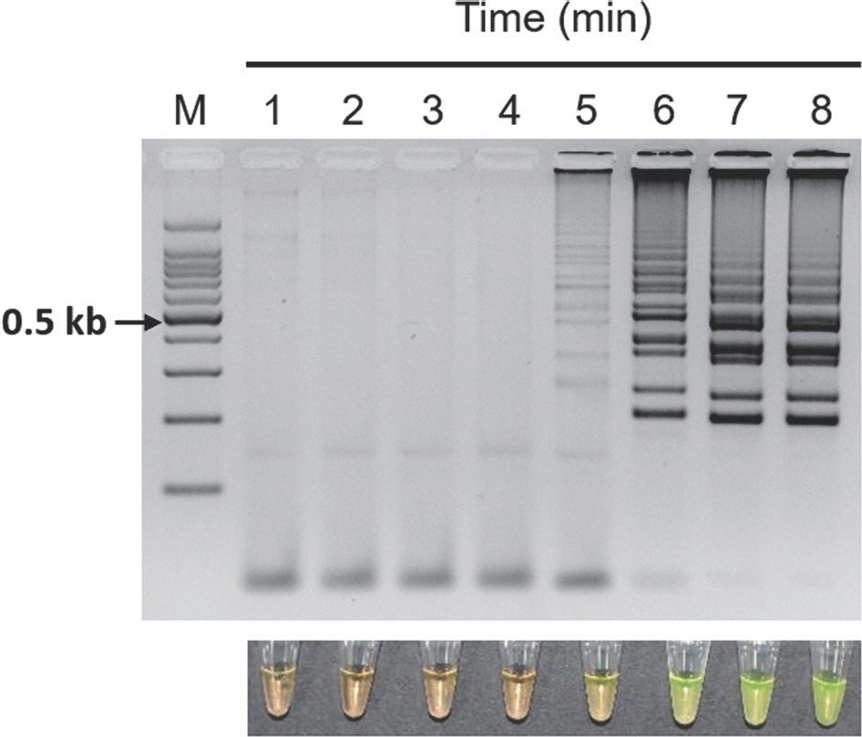
Effect of reverse transcription loop-mediated isothermal amplification (RT-LAMP) incubation time on the detection of Apple stem grooving virus (ASGV). RT-LAMP results were evaluated by 2% agarose electrophoresis and SYBR Green I. Orange color represents negative and yellow color represents positive. Lane M, 100-bp DNA ladder; lanes 1–8, 1, 5, 10, 20, 30, 40, 50, and 60 min, respectively.
Specificity of RT-LAMP assay. The specificity of the RT-LAMP assay for ASGV was evaluated against ACLSV and ASPV, which are also major pathogens of pear trees in Korea. The RT-LAMP assay generated amplification in ASGV-infected samples and yielded no amplification from either the ACLSV-or ASPV-infected samples (Fig. 3). The RT-PCR assays confirmed the presence of the viruses in each of virus-infected pear leaf sample. As a result of the reaction, no amplification was observed for either type of negative control (i.e., healthy plant samples and non-template control). Thus, the RT-LAMP assay was determined to be a reliable method for ASGV detection.

Specificity evaluation of reverse transcription loop-mediated isothermal amplification (RT-LAMP) assay for Apple stem grooving virus (ASGV) detection. RT-LAMP results were evaluated by 2% agarose electrophoresis and SYBR Green I. Orange color represents negative and yellow color represents positive. nad5 was used as a housekeeping gene. RT-PCR, reverse transcription polymerase chain reaction. Lane M, 100-bp DNA ladder; lane 1, ASGV-infected pear leaf sample (positive control); lane 2, apple chlorotic leaf spot virus (ACLSV)-infected pear leaf sample; lane 3, apple stem pitting virus (ASPV)-infected pear leaf sample; lane 4, healthy pear leaf sample (negative control); lane 5, non-template.
Sensitivity of RT-LAMP assay. The sensitivity of RT-LAMP and RT-PCR was evaluated using serially diluted total RNA from ASGV-infected leaf samples. The detection limit of the RT-LAMP assay for ASGV detection was 1.5×10⁻⁶ μg, whereas RT-PCR yielded very weak amplification at 1.5×10⁻⁵ and 1.5×10⁻⁶ μg template RNA (Fig. 4). Therefore, the RT-LAMP assay for ASGV detection confirmed ~100-fold higher sensitivity than the conventional RT-PCR assay.
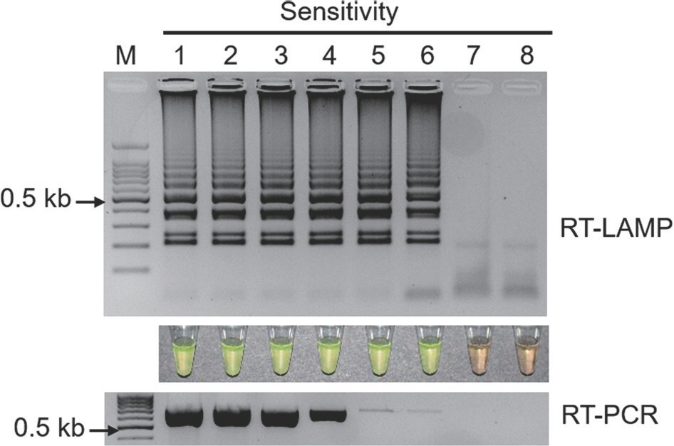
Sensitivity evaluation of reverse transcription loop-mediated isothermal amplification (RT-LAMP) and reverse transcription polymerase chain reaction (RT-PCR) for Apple stem grooving virus (ASGV) detection. RT-LAMP results were evaluated by 2% agarose electrophoresis and SYBR Green I. Orange color represents negative and yellow color represents positive. RT-PCR results were loaded in 2% agarose gel. Lane M, 100-bp DNA ladder; lanes 1–8, 1.5×10⁻¹, 1.5×10⁻², 1.5×10⁻³, 1.5×10⁻⁴, 1.5×10⁻⁵, 1.5×10⁻⁶, 1.5×10⁻⁷, and 1.5×10⁻⁸ dilutions of total RNA.
Performance of RT-LAMP using field-collected samples. The reliability of the optimized RT-LAMP assay was evaluated using eight field-collected and symptom-free pear leaf samples from major local pear orchards in Sangju, Naju, Ulsan, and Cheonan, Korea. The RT-LAMP assay detected ASGV in all collected samples but not in healthy plants (negative controls). However, seven tested samples were positive to RT-PCR, and one tested sample showed weak amplification, which indicated the relatively higher sensitivity of the RT-LAMP assay, when compared to conventional RT-PCR (Fig. 5). These results demonstrated the superior reliability of RT-LAMP for ASGV detection in field-collected samples.
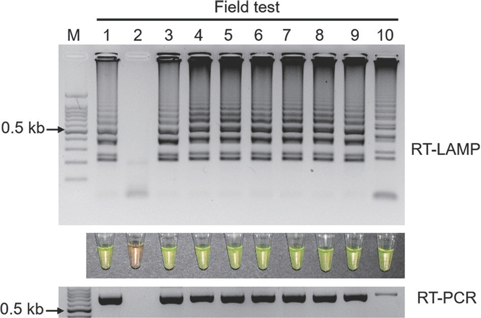
Sample test of reverse transcription loop-mediated isothermal amplification (RT-LAMP) for Apple stem grooving virus (ASGV) detection. The RT-LAMP results were evaluated by 2% agarose electrophoresis and SYBR Green I. Orange color represents negative and yellow color represents positive. Lane M, 100-bp DNA ladder; lane 1, ASGV-infected pear leaf sample (positive control); lane 2, healthy pear leaf sample (negative control); lanes 3–4, pear leaf samples collected from Sangju; lane 5, pear leaf sample collected from Naju; lanes 6–7, pear leaf samples collected from Ulsan; lanes 8–10, pear leaf samples collected from Cheonan.
Paper-based RT-LAMP. Even though the RT-LAMP assay was successfully detected ASGV in pear leaf samples, the main purpose for developing the RT-LAMP assay was to apply it under field conditions in combination with simple, on-site RNA extraction. Thus, RT-LAMP assay was performed and evaluated using RNA extracted using a paper-based method. Both the RT-LAMP and RT-PCR assays were able to detect ASGV in pear samples isolated using paper-based methods and failed to detect viral RNA in healthy leaf samples (Fig. 6). Therefore, the paper-based RT-LAMP assay could be used for the detection of ASGV under field conditions.
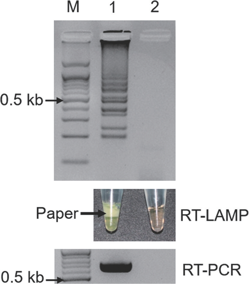
Evaluation of paper-based RNA isolation for the reverse transcription loop-mediated isothermal amplification (RT-LAMP)-mediated detection of Apple stem grooving virus (ASGV). Whatman No. 1 discs were used to isolate RNA from pear leaf samples and added to RT-LAMP reactions. The RT-LAMP results were visualized by 2% agarose electrophoresis and SYBR Green I. Orange color represents negative and yellow color represents positive. The products of reverse transcription polymerase chain reaction (RT-PCR) were loaded in 1.2% agarose gel. Lane M, 100-bp DNA ladder; lane 1, ASGV-infected pear leaf sample (positive control); lane 2, healthy pear leaf sample (negative control).
Discussion
ASGV is a common virus among economically important fruit trees, including apple and pear (Canales et al., 2021). A recent survey study suggested that the incidence of ASGV in pear production in Korea was >90% (Kim et al., 2019); besides, ASGV has also been classified as a high-risk viral pathogen to produce virus-free apple and pear germplasm and plantlets by Korea Seed and Variety Service (Lee et al., 2017). Thus, a rapid, simple, and sensitive detection method is urgently required to prevent the further spread of ASGV.
Although several diagnostic methods have been developed for ASGV detection, these methods generally require specialized equipment, well-trained personnel, and relatively long reaction time (James, 1999; Yao et al., 2014). A RT-LAMP assay has also been studied and reported for ASGV detection in apple leaf samples in China; however, the assay used primers that were only based on genome sequences from a few Chinese ASGV isolates, and the specificity and field performance of the assay have not been reported (Zhao et al., 2014). In this study, a simple, specific, sensitive, and reliable new RT-LAMP assay was developed for the detection of ASGV. The RT-LAMP assay utilized SYBR Green I, which facilitates the visual confirmation of amplification via visually discernable color changes, thereby negating the necessity for gel electrophoresis and evaluation (Notomi et al., 2000). The optimized one-step RT-LAMP assay was also able to detect ASGV in RNAs from ASGV-infected leaf samples at 63°C for 50 min using an isothermal device such as a water bath, whereas conventional RT-PCR required at 2–3 hr using a thermocycler to detect ASGV, and the RT-LAMP assay had higher specificity and 100-fold higher sensitivity compared to that of conventional RT-PCR assay. The reaction time of 50 min required to detect ASGV by the RT-LAMP assay was similar to 50–60 min observed in RT-LAMP studies detecting other plant viruses (Lee and Jeong, 2020; Lu et al., 2018). Moreover, the RT-LAMP assay was validated using field-collected samples from pear orchards and was compatible with paper-based RNA isolation.
Molecular diagnostics under field conditions is often hampered by nucleic acid isolation, which can be difficult and laborious without typical molecular biology equipment (Zou et al., 2017). Paper-based RNA isolation methods convert isothermal amplification assays, including RT-LAMP, to quick and simple diagnostic applications that can be performed outside the laboratory. Thus, we plan to use the combined paper-based isolation and RT-LAMP strategy for a nation-wide ASGV survey.
Taken together, this study describes an optimized RT-LAMP assay as a quick visual method for the rapid, specific, sensitive, and reliable detection of ASGV in pear leaves. The optimized assay requires only heating equipment and, therefore, it can be easily implemented in field conditions. Considering its advantages, the technique is promising for the rapid and extensive detection of ASGV in the field and in virus-free certification programs.
Notes
Conflicts of Interest
No potential conflict of interest relevant to this article was reported.
Acknowledgments
This work supported by Korea Institute of Planning and Evaluation for Technology in Food, Agriculture, Forestry and Fisheries (IPET) through (Agri-Bioindustry Technology Development Program), funded by Ministry of Agriculture, Food and Rural Affairs (MAFRA) (No. 317006-04-2-HD030).
Electronic Supplementary Material
Supplementary materials are available at Research in Plant Disease website (http://www.online-rpd.org/).

