Colletotrichum fioriniae에 의한 호박 과실 탄저병의 발생 보고
First Report of Colletotrichum fioriniae Causing Anthracnose on Fruit of Pumpkin (Cucurbita moschata) in Korea
Article information
Abstract
In 2014, Anthracnose symptoms were observed on fruit of pumpkin in Gangneung, Gangwon in Korea. The typical symptoms on fruit surfaces were circular lesions, brown, shriveled with salmon-colored spore masses on the spots. A fungal isolate was obtained from symptomatic on fruit and cultured on potato dextrose agar at 25°. The color of the upper surface of the colony varied from white to gray with occurrence of salmoncolored conidial masses and was red to violet on the underside. The causal fungus of anthracnose isolated from the diseased fruits was identified as Colletotrichum fioriniae based on mycological characteristics and sequences of the internal transcribed spacer, glyceraldehyde-3-phosphate dehydrogenase, chitin synthase-1 gene, histone H3, actin gene, beta-tubulin gene region. Pathogenicity tests of the isolate were performed on pumpkin fruit by both wound and non-wound inoculation with conidial suspension (1.106 conidia/ml) and incubated at 25°. Lesions were observed on both wounded and non-wounded fruit of pumpkin after 7 days inoculation in only C. fioriniae. The pathogen was re-isolated from the lesions of inoculated fruits. To our knowledge, this is the first report of anthracnose on fruit of pumpkin caused by C. fioriniae in Korea
박과 작물에 속하는 호박은 1년생 덩굴성 초본으로 열대아메리카가 원산지이며, 종류와 특성에 따라 동양계 호박(Cucurbita moschata Duch.), 서양계 호박(C. maxima Duch.), 폐포계 호박(C. pepo L.) 등으로 구분된다. 호박의 노지, 시설재배에서 국내 생산면적은 각각 9,206 ha, 2,097 ha이며, 생산량은 각각 310,218톤, 138,166톤으로 과채류 중에서 가장 많은 부분을 차지하고 있다(Statistics Korea, 2018). 호박은 품종과 숙성기간에 따라 효능과 영양소가 달라 식품적 가치가 높고, 병충해가 적고 재배가 용이하여 전국 어디에서나 재배가 가능하다.
국내에서 호박에 가해하는 병은 곰팡이 20종, 바이러스 8종, 세균 2종, 선충 1종 총 31종이 보고되어 있다(Korean Society of Plant Pathology, 2009). 그 중 Colletotrichum orbiculare (=Colletotrichum lagenarium)에 의한 탄저병은 호박을 포함한 박과 작물에 발생하며, 생육기 전반 걸쳐 잎, 줄기, 과실에 발생하고, 수확 후 저장 중에 발생하여 무름병을 일으킨다.
2014년 강원도 강릉시 사천면 시설하우스에서 재배중인 호박 과실에 탄저병 증상이 처음으로 관찰되었다. 호박 과실에 발병한 탄저병은 기존에 보고된 C. orbiculare에 의한 탄저병과 차이가 있었으며, 과실에 분홍색의 분생포자층이 동심원을 그리면서 진전됨에 따라 병반이 확대되어 결국 과실이 무르는 증상이 나타났다(Fig. 1A, B). 병든 과실로부터 원인균을 규명하기 위해 순수 분리를 하여 균주를 획득하였다. 병원균의 균학적 특성, 병원성 검정, 염기서열 분석을 통해 호박 과실 탄저병의 원인균을 규명하였다.
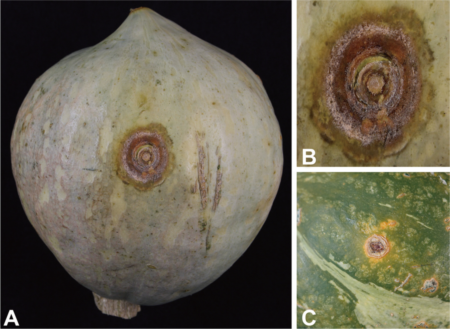
Symptoms of anthracnose on fruit of pumpkin caused by Colletotrichum fioriniae. (A) Naturally occurred symptom. (B) Close-up of infected fruit surface. (C) Symptoms on an artificially inoculated fruit.
병원균의 분리
호박 과실에 발달된 탄저병의 분생포자층을 멸균된 백금이를 이용하여 1 loop 취한 후, streptomycin(100 μg/ml)이 함유된 potato dextrose agar (PDA) 배지에 획선평판법(streaking plate) 방법을 이용하여 단포자를 분리하였다. 25°에서 3일간 배양하여 자란 균사의 선단 부분을 취해 새로운 PDA 배지에 배양하였다. 순수 분리된 대표균주를 농업유전자원센터에 기탁하였다(KACC47696).
병원균의 균학적 특성
탄저병 증상을 보이는 호박 과실로부터 분리된 대표균주를 균학적 및 형태학적 특징을 관찰하기 위해서 PDA 배지에 25°에서 10일간 배양하였다. PDA 배지상에서의 균총은 하얀색에서 회백색을 띠었고, 오렌지색의 분생포자층을 형성하였다. 분생포자는 투명하고 격벽이 없으며, 양쪽 끝이 약간 뾰족한 방추형이거나 원통형으로 크기는 8.3–14.9×2.9–4.3 μm이다. 부착기는 옅은 갈색의 넓은 타원형 내지 곤봉형 이거나 불규칙한 모양이며, 크기는 7.3–11.5×4.8–7.4 μm이다. 이러한 병원균의 균학적 특성은 C. fioriniae와 일치하였다(Shivas와 Tan, 2009). C. fioriniae는 C. acutatum의 종 복합체로 알려져 있다(Sreenviasaprasad와 Talhinhas, 2005). 그러나 Shivas와 Tan (2009)에 의해 다른 종들과 명확하게 분리되는 종으로 규명되었다(Table 1, Fig. 2A–E).

Comparison of mycological characteristic of present fungus isolated from fruit of pumpkin
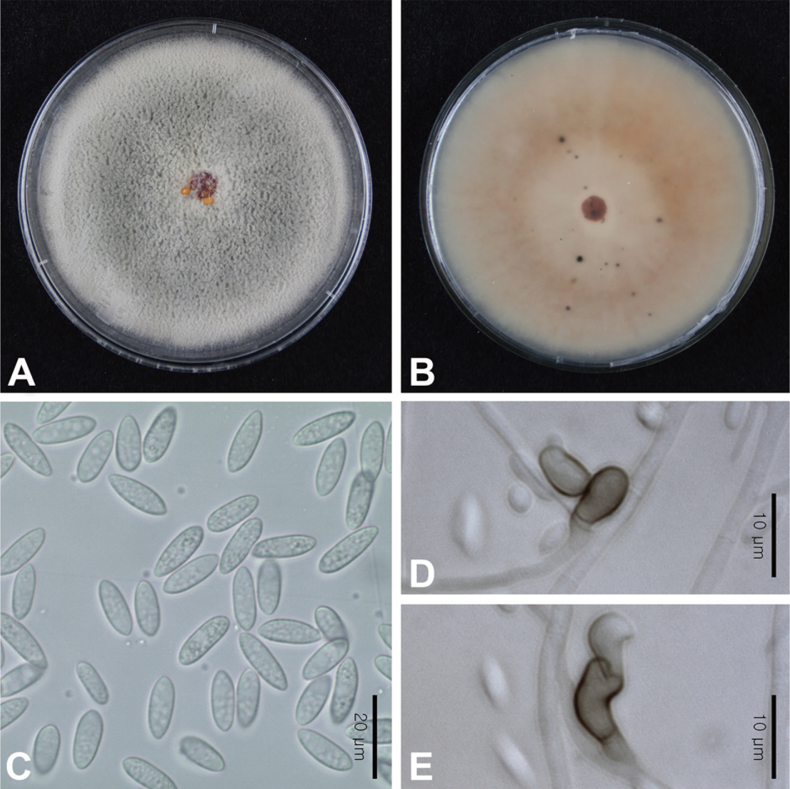
Morphological characteristics of Colletotrichum fioriniae isolated from fruit of pumpkin. (A) Upper surface of colony incubated on potato dextrose agar (PDA). (B) Underside surface on PDA. (C) Conidia. (D, E) Appressoria. Scale bars=20 μm (C), 10 μm (D, E).
병원성 검정
분리된 대표균주를 PDA 배지에 치상하여 25°에서 10일 동안 배양 후, 형성된 포자를 수확하여 멸균수를 이용하여 1.0×10⁶ conidia/ml로 조정하여 현탁액을 제조하였다. 병원성 검정을 위하여 신선한 호박 과실을 70% 에탄올과 1% NaOCl을 이용하여 표면 살균하였고, 멸균수를 이용하여 2회 씻은 후 건조하여 사용하였다. 접종 방법은 멸균된 needle을 이용하여 상처를 유도한 방법과 상처를 내지 않은 방법을 이용하였으며, 병원균 포자 현탁액을 각각의 처리구에 10 μl씩 pipetting하였고, 무처리구에는 멸균수를 이용하여 같은 방법으로 접종하여 고습도 습실상(95% relative humidity, 25°)에서 발병을 유도하였다. 접종 7일차 모든 접종원에서 멸균수를 처리한 무처리구를 제외한 호박 과실에서 처음 발병한 호박 과실탄저병과 유사한 증상을 보였다. 병든 과실을 재분리 결과 동일한 균이 반복적으로 재분리 되었다(Fig. 1C).
계통학적 유연관계 분석
호박 과실로부터 분리된 대표균주의 형태적 특성을 바탕으로 명확한 종 동정을 위해 염기서열 분석을 수행하였다. 대표균주를 potato dextrose broth 배지에서 25°에서 5일간 180 rpm으로 배양한 후, 균체를 걸러 액체질소를 이용하여 마쇄한 후 DNeasy Plant Mini Kit (Qiagen, Hilden, Germany)를 이용하여 genomic DNA를 획득하였다. PCR를 수행하기 위해 internal transcribed spacers and intervening 5.8S rDNA (ITS), partial glyceraldehyde-3-phosphate dehydrogenase (GAPDH), partial chitin synthase-1 gene (CHS-1), partial histone H3 (HIS3), partial actin gene (ACT), partial β-tubulin gene (TUB2) 영역을 증폭하였다. ITS 영역은 ITS 1/ITS 4 (White 등, 1990), GAPDH 영역은 GDF/GDR (Templeton 등, 1992), CHS-1 영역은 CHS-354R/CHS-79F (Carbone과 Kohn, 1999), HIS3 영역은 CYLH3F/CYLH3R (Crous 등, 2004), ACT 영역은 ACT-512F/ACT-783R (Carbone과 Kohn, 1999), TUB2 영역은 T1/Bt2b (O’Donnell과 Cigelnik, 1997; Glass and Donaldson, 1995) 프라이머를 사용하였다. 각각 영역의 증폭된 PCR 산물을 정제한 후, 염기서열을 분석하기 위해 Macrogen Co. Ltd. (Seoul, Korea)에 의뢰하였고, SeqMan (DNASTAR, Madison, WI, USA)과 CLUSTRAL W을 이용하여 정렬하였다. 분석된 염기서열은 NCBI (National Center for Biotechnology Information) BLAST search를 통하여 상동성을 비교 결과, ACT 영역에서 C. fioriniae 균주와 99% 이상 일치하였고(LC516662), 나머지 영역에서는 C. fioriniae 균주와 100% 일치하였다(KY695261, MN895544, MN974140, JQ949284, AB744086). 대표 균주는 NCBI GenBank에 등록하였다(accession nos. KT728897, MT978180, KT728900, MT978181, KT728898, KT728899). 계통수를 작성하기 위해 분석된 염기서열의 ITS, GAPDH, CHS-1, HIS3, ACT, TUB2 각각의 영역을 연결하여, MEGA 7을 이용하여 maximum likelihood 방법으로 작성하였고, 1,000회 bootstrap을 수행하였다. Reference sequence 로는 C. acutatum의 종 복합체인 C. scovillei, C. nymphaeae, C. simmondsii, C. lupine, C. melonis, C. fioriniae, C. acutatum, C. acerbum, C. salicis, C. godetiae, C. pyricola 균주와 outgroup으로는 C. truncatum, C. gloeosporioides을 사용하였다. 계통수 분석 결과, 대표균주 KACC47696은 C. acutatum의 종 복합체 clade에 속하였으며, C. fioriniae (CBS127600)와 묶여 다른 종들 과 명확하게 구분되었다(Fig. 3.)
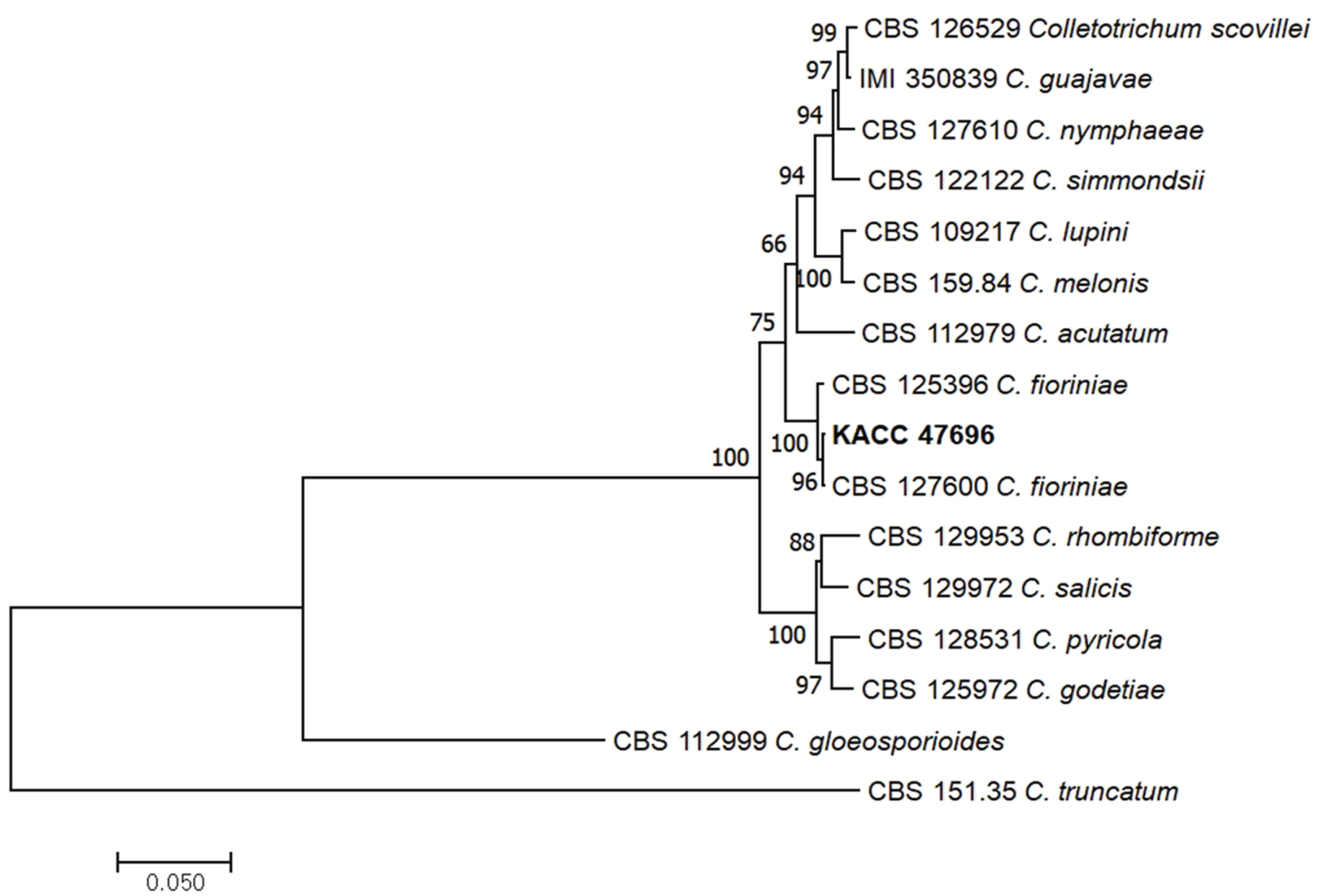
Phylogenetic tree inferred from maximum likelihood analysis derived from sequence of combined multi-locus gene (ITS, GAPDH, CHS-1, HIS3, ACT, and TUB2) of Colletotrichum fioriniae and those of other Colletotrichum spp. The numbers above the nodes are supporting percentages by 1,000 bootstrap replicates.
C. fioriniae 종은 C. acutatum의 종 복합체로 유전적으로 다른 종들과 명확하게 구별되는 종으로 Clade 3에 속하며 (Damm 등, 2012), Shivas와 Tan (2009)에 의해서 규명되었다. 국내에서 C. fioriniae에 의한 탄저병의 보고는 구기자나무, 복숭아나무, 자두나무, 가지 등에서 보고되었으며(Hassan 등, 2019; Lee 등, 2018; Oo 등, 2016; Xu 등, 2018), 박과 작물에서는 보고가 되어 있지 않다. 또한, 국내에서 호박에 발생하는 탄저병은 C. orbiculare가 보고되어 있으나, C. fioriniae에 의한 호박 과실 탄저병은 아직까지 보고된 바 없다. 따라서 본 연구는 국내에서 처음으로 C. fioriniae에 의한 호박 과실 탄저병의 발생을 보고하고자 한다.
요 약
강원도 강릉시 사천면 비닐하우스에서 재배중인 호박 과실에 탄저병이 발생하였다. 병든 호박 과실에 분홍색의 분생포자층이 동심원으로 나타나 점차 확대되어 과실이 무르는 증상을 나타내었다. 원인균을 규명하기 위하여 순수 분리 후 균학적 특성 및 ITS, GAPDH, CHS-1, HIS3, ACT, TUB2 염기서열 분석결과 Colletotrichum fioriniae로 동정하였다. 또한, 병원성이 확인되었고 접종시험에서 동일한 균이 반복적으로 분리되었다. 따라서, 이러한 결과를 바탕으로 C. fioriniae에 의한 호박 과실에 발생하는 탄저병의 발생을 국내 처음으로 보고한다.
Notes
Conflicts of Interest
No potential conflict of interest relevant to this article was reported.