

서론
아까시나무의 원산지는 북아메리카이며 낙엽활엽교목이다. 일제강점기에 황폐화된 산지의 녹화와 연료림을 목적으로 우리나라에 식재가 권장되었다고 한다(Lim, 1994). 아까시나무는 조경수, 사방지 녹화수, 꿀을 제공하는 밀원식물로 쓰인다. 꽃은 향료로도 쓰이고 잎은 식용하기도 하고, 수피는 제지, 섬유, 목재로도 쓰인다(Lee 등, 2010). 우리나라에 자생하는 아까시나무의 병해는 모자이크병, 빗자루병 등을 비롯한 총 12종의 병해가 보고되어 있다(The Korean Society of Plant Pathology, 2009). 2014년 8월 안동대학교 인근 야산 및 청송, 문경의 야산에 자생하는 아까시나무 잎에 진한 갈색 혹은 검은 반점이 형성되고, 잎이 황화되는 것을 관찰하였다. 병든 식물체를 채집하여 병원균을 순수 분리한 뒤 균학적 특성과 병원성 검정을 하였으며, internal transcribed spacer (ITS) ribosomal RNA 유전자 염기서열을 분석하여 원인병원균을 동정하였다. 그 결과 분리된 병원균은 Colletotrichum acutatum으로 동정하였다. 우리나라에서 C. gloeosporioides에 의한 아까시탄저병은 보고된 바 있으나, C. acutatum에 의한 아까시나무 탄저병은 보고된 적이 없다. 따라서 본 연구에서는 아까시나무에 발병한 탄저병의 병징, 병원성 검정, 균학적 특성 및 ITS 염기서열 분석 결과를 보고한다.
병원균 분리 및 균학적 특성
아까시나무에서 발병한 검은 반점 및 황변한 이병엽의 병원균을 분리하기 위하여 안동시, 청송군, 문경시 소재 야산 20여 개 장소에서 이병식물체를 채집하였다(Table 1). 병원균을 분리하기 위해 채집한 아까시나무 잎의 이병 부위를 건전부와 이병부가 반씩 되도록 작게 잘랐으며, 각 시료를 70% 에탄올과 1% NaOCl 용액에 30초간 짧은 시간 표면소독 후 멸균수에 2회 세척하였다. 세척 후 멸균된 필터 페이퍼를 이용하여 물기를 완전히 제거하고 5분 정도 자연 건조시킨 후, potato dextrose agar (PDA; Difco, Detroit, MI, USA)에 치상하여 28°C 암조건에서 배양하였다. 배양 3-5일 후, 각 시료에서 분리된 곰팡이의 콜로니 형태와 색깔을 관찰하였으며, 관찰결과 균총의 색은 흰색으로 동일하였다. 자라나온 곰팡이의 선단을 메스로 잘라내어 새로운 PDA의 중앙에 치상하고, 10일 배양 후 petri dish에 가득 자란 곰팡이의 분생포자를 현미경으로 관찰하였다. 포자현탁액의 일부를 PDA에 도말하여 1일 배양 후 광학현미경을 보면서 단포자(single spore isolation) 분리하여 이후 실험에 사용하였다. 분리된 곰팡이는 광학현미경(BX43; Olympus, Tokyo, Japan)을 이용하여 포자의 형태, 크기 및 부착기 형태, 색깔, 크기 등의 균학적 특성을 관찰하였다. 포자와 부착기의 크기는 현미경에 부착된 image analyzer (ProgRes® Speedxtcore3; Jenoptik, Jena, Germany)를 이용하여 측정하였다. PDA 배지상에서 균총은 흰색을 띠었으나 점차적으로 회색으로 변하였고, 오렌지색의 분생포자층을 형성하였다(Fig. 2A). 균사는 격막이 있고 투명하였으며, 분생포자는 끝이 약간 뾰족한 방추형의 형태를 띠었다. 분생포자의 크기는 평균 길이 8.3-17.2 μm, 너비 2.5-4.1 μm였다(Fig. 2B). 부착기는 암갈색으로 곤봉상이며, 크기는 8.1-12.3×4.1-6.4 μm였다(Fig. 2C, Fig. 2 Table 2) (Damm 등, 2012; Sutton, 1980).
Table 1
Isolation of Colletotrichum acutatum fungal pathogens from the symptomatic tissues of black locust (Robinia pseudoacacia) obtained from various locations in three different cities in Korea
Fig. 2
Morphological characteristics of Colletotrichum acutatum. (A) Colony morphology on potato dextrose agar after 10 days of incubation. (B, C) Conidia and appressoria formation.
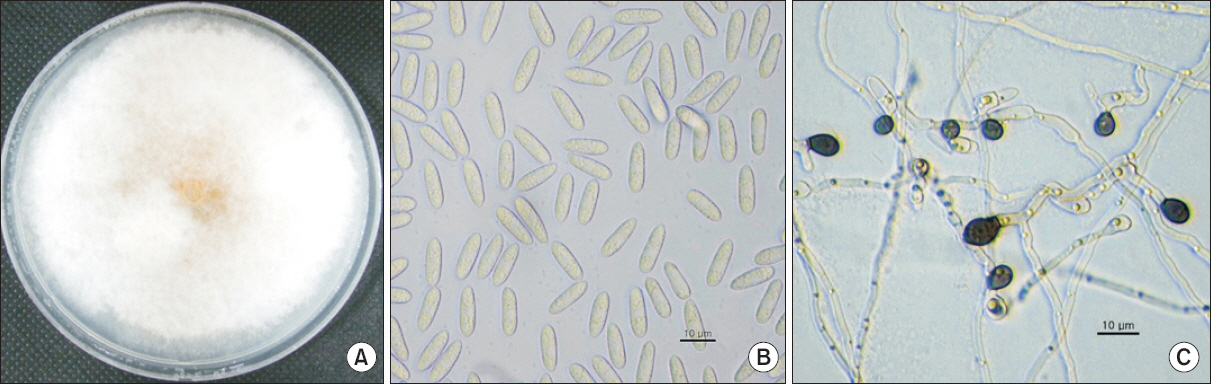
Table 2
Comparison of morphological characteristics of the fungus isolated from black locust (Robinia pseudoacacia) with those of Colletotrichum acutatum described previously
| Characteristic | Present isolate | C. acutatum* | C. acutatum† | |
|---|---|---|---|---|
| Colony | Pinkish gray | Smoke gray to olivaceous gray | White to pinkish gray | |
| Conidia | Shape | Straight, fusiform | Aseptate, straight, cylindrical to fusiform | Fusiform |
| Size (µm) | 8.3-17.2×2.5-4.1 | 7.5-19×3.5-4.5 | 8.5-16.5×2.5-4 | |
| Appressoria | Color | Pale to dark brown | Medium brown | Pale to dark brown |
| Shape | Clavate, obovate | Ellipsoidal to obovate, undulate | Clavate | |
| Size (µm) | 8.1-12.3×4.1-6.4 | 4-13×4-6.5 | 8.5-10×4.5-6 | |
병원성 검정
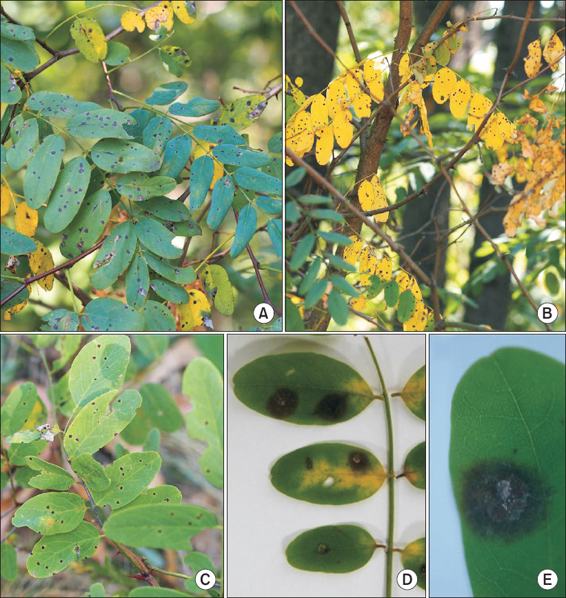
아까시나무 잎에서 분리한 병원균의 병원성을 확인하기 위하여, 야외에서 자라는 건전한 아까시나무 가지를 채집하여 사용하였다. 병원성 검정 균주는 안동대학교 야산에서 분리한 균주(A3)를 사용하였다. 잎의 표면에 채혈기를 이용하여 상처를 낸 뒤 25°C에서 10일간 배양한 곰팡이의 포자 현탁액(1×10⁵ conidia/ml)을 10 μl씩 상처 부위에 떨어뜨려 접종하였다. 접종한 아까시나무 잎은 플라스틱 용기(20×15×8 cm)에 담아 포화습도를 유지한 후, 25°C growth chamber에 두고 발병유무를 관찰하였다. 3반복으로 2회 재현실험을 수행하였다. 접종 2-3일 후, 잎에 병원균을 접종한 부위에서 검은 반점이 형성되기 시작하였고, 시간이 지날수록 병반이 점차 확대되었다. 5-7일 후부터는 병반 부위에 하얀 곰팡이 균사가 형성되는 것을 확인하였다(Fig. 1D, Fig. 1E). 또한 무상처 병원성 검정을 위해 야외에서 자라고 있는 아까시나무를 화분에 옮겨 심어 30일 동안 온실에서 키웠다. 잎의 표면을 살균수로 세척하고, 병원균 현탁액 (1×10⁵ conidia/ml) 100 ml를 흘러 떨어지도록 충분히 분무 접종하였다. 접종한 가지는 비닐봉지를 씌워 습도를 유지시켰으며 22°C-27°C 온실에 두었다. 48시간 후 비닐 봉지를 벗기고 온실에 두면서 발병유무를 조사하였다. 접종 5-7일 후부터 병원균을 접종한 잎에 반점이 보이기 시작하였고, 소형 반점은 점점 확대되어 2-3주가 경과하였을 때 직경 1-2 mm 정도의 반점을 형성하였다. 일부 병징 주변에는 노란 달무리가 형성되기도 하였다(Fig. 1C). 자연상태에서 상처를 내지 않았을 때 병 발생은 실내에서 상처를 내었을 때보다 많은 시간이 요구되었다. 아까시나무 잎에서 분리한 병원균은 상처 및 무상처 접종을 통해 병원균의 병원성을 확인하였으며, 자연상태에서와 유사한 병징을 나타내었다. 재현된 병징에서 병원균을 분리하여 균학적 특성 및 염기서열 분석 결과 분리하여 접종한 병원균과 동일한 특성을 나타내었다.
ITS 염기서열분석
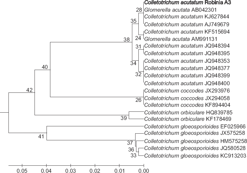
아까시나무 잎에서 분리한 병원균의 동정을 위해 ITS region의 염기서열을 분석하였다. 각 지역에서 분리된 21균주를 분석하였다. White 등(1990)이 사용한 primers ITS1 (5’-TCCGTAGGTGAACCTGCGG-3’)과 ITS4 (5’-TCCTCCGCTTATTGATATGC-3’)를 이용하여 증폭하였으며 증폭된 PCR 산물은 NucleoSpin Gel과 PCR Clean-up Kit (Macherey-Nagel, Düren, Germany)를 사용하여 순화하였다. 염기서열 분석은 SolGent (Daejeon, Korea)에 의뢰하였으며, 분석된 21균주의 염기서열을 서로 비교해 본 결과 ITS 염기서열이 동일하였다. 그 중 한 균주(안동대학교 인근 야산에서 분리된 균주 A3)의 염기서열을 National Center for Biotechnology Information (NCBI)의 BLASTN을 이용하여 GenBank에 등록되어 있는 균주들과 비교하였으며, 염기서열은 GenBank에 등록하였다(accession No. KU881799). 아까시나무에서 분리한 C. acutatum의 ITS 염기서열은 다른 기주에서 탄저병을 일으키는 C. acutatum의 ITS 염기서열(accession Nos. KJ627844, AJ749679)과 100% 일치하였다(Fig. 3).
Fig. 3
Phylogenetic relationships among Colletotrichum acutatum isolates, including a anthracnose infecting black locust, based on a similarity analysis of internal transcribed spacer (ITS) sequence.
이상과 같이 병원균의 균학적 특성, 병원성검정 및 ITS 염기서열 분석 결과 C. acutatum으로 동정되었다. 이는 고추 탄저병(Han 등, 2009), 배암차즈기 탄저병(Kwon 등, 2007)에 보고된 병원균과 균학적 특징이 일치하였다. 우리나라에서 C. acutatum이 병원균으로 보고된 기주는 석류나무, 녹두, 고추, 배나무, 복숭아, 사과, 포도나무, 구기자나무, 잇꽃, 베고니아, 코스모스, 배암차즈기이다. 이에 C. acutatum에 의한 아까시나무 탄저병을 새로이 보고한다. 아까시나무 탄저병은 병징이 여러 가지로 나타나는 것을 확인하였다. 소형 검은 반점이 형성되는 병징 이외에 반점이 없이 황화되어 낙엽되는 병징, 잎의 가장자리가 황변되며 말리는 병징이 관찰되었다. 병징에 따른 탄저병균의 특성과 병 발생기작에 대한 연구가 동반되어야 할 것으로 생각된다. 최근 고추와 사과에 발병하는 Colletotrichum spp.에 대한 살균제 저항성에 따른 유전적 차이(Kim 등, 2014) 및 생물학적 방제(Han 등, 2015; Hong 등, 2015)에 관한 연구들이 많이 이루어지고 있다. 그러므로 아까시나무에서 분리되어지는 탄저병균(C. gloeosporioides 및 C. acutatum)과 주요 경제작물인 사과나무, 고추 등과의 전염 가능성 및 병원성과의 상관관계는 더 연구할 필요성이 있을 것으로 생각된다.
 PDF Links
 PubReader
 Full text via DOI
 Download Citation
 Print
Print


