

ņä£ļĪĀ
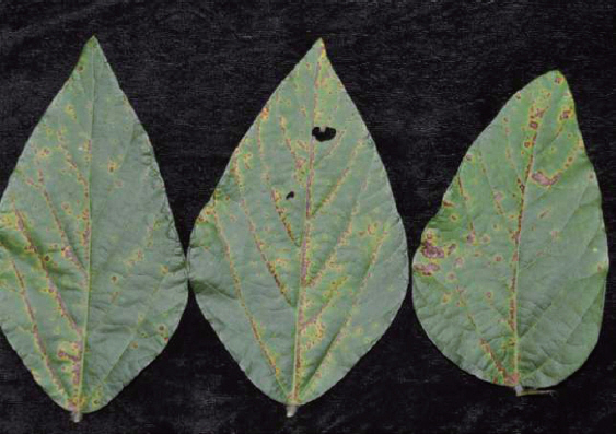
ņĮ®ņØĆ ņäĖĻ│äņĀüņ£╝ļĪ£ ņżæņÜöĒĢ£ ņŗØļ¤ēņ×æļ¼╝ņØ┤ļ®░ ĻĄŁļé┤ņŚÉņä£ļŖö ļ®öņŻ╝ņØś ņøÉļŻīļĪ£ ņé¼ņÜ®ļÉśņ¢┤ ņÜ░ļ”¼ļéśļØ╝ ņŗØņāØĒÖ£ņŚÉ ļ░śļō£ņŗ£ ĒĢäņÜöĒĢ£ ņ×æļ¼╝ņØ┤ļŗż. ņĮ®ņŚÉ ļ│æņØä ņØ╝ņ£╝ĒéżļŖö ļ│æņøÉņ▓┤ļŖö ļ░öņØ┤ļ¤¼ņŖż, ņ¦äĻĘĀ, ņäĖĻĘĀ ļō▒ņ£╝ļĪ£ ņØ┤ ņżæ ņäĖĻĘĀņŚÉ ņØśĒĢ£ ļ│æņØĆ ļČłļ¦łļ”äļ│æ, ņäĖĻĘĀņĀÉļ¼┤ļŖ¼ļ│æ, ņäĖĻĘĀĻ░łņāēņĀÉļ¼┤ļŖ¼ļ│æ(KSPP, 2009), ļōżļČłļ│æ(Myung ļō▒, 2009)ņØś 4ņóģņØ┤ ļ│┤Ļ│ĀļÉśņ¢┤ ņ׳ļŗż. ļČłļ¦łļ”äļ│æņØĆ Xanthomonas axonopodis pv. glycinesņŚÉ ņØśĒĢ┤ ļ░£ņāØĒĢśļŖöļŹ░ ņĀäĒśĢņĀüņØĖ ļ│æņ¦ĢņØĆ ņŻ╝ļ│ĆņØ┤ ņśģņØĆ ĒÖ®ņāēņØĖ ņĀüĻ░łņāēņØś ņøÉĒśĢļ│æļ░śņ£╝ļĪ£ ņ×Ä ņĀäļ®┤ņŚÉ ļ│æļ░śņØ┤ ļéśĒāĆļéśļ®░ ņŻ╝ļĪ£ ņŚĮļ¦źņØä ņżæņŗ¼ņ£╝ļĪ£ ļ│æļ░śņØ┤ ļČäĒżĒĢ£ļŗż(Fig. 1). ļ│æņ¦Ģ ļśÉļŖö ļ│æņøÉĻĘĀņØś ļČäĒżņŚÉ Ļ┤ĆĒĢ£ ņŚ░ĻĄ¼ļĪ£ ņśżņØ┤ ņ×ÄņŚÉņä£ Pseudomonas syringae pv. lachrymansņØś ņāüļīĆ ņŖĄļÅäņŚÉ ļö░ļźĖ ļČäĒżĻ░Ć ņ×Ä ĻĖ░ļČĆņÖĆ ņŚĮļ¦źņŻ╝ņ£äļĪ£ ļČäĒżĒĢ£ļŗżĻ│Ā ļ│┤Ļ│ĀĒĢśņśĆņ£╝ļ®░(Leben, 1988), Krimm ļō▒(2005)ņØĆ ĒśĢĻ┤æĒśäļ»ĖĻ▓ĮņØä ņØ┤ņÜ®ĒĢśņŚ¼ ļöĖĻĖ░ņØś trichomeņŚÉ ņäĖĻĘĀņØ┤ ļČĆņ░®ļÉ£ Ļ▓āņØä Ļ┤Ćņ░░ĒĢśņŚ¼ ļ│┤Ļ│ĀĒĢ£ ļ░ö ņ׳ļŗż. ļśÉĒĢ£ ņäĖĻĘĀņØś ņ╣©ņ×ģņØĆ ĻĖ░Ļ│ĄņØś Ļ░£ĒÅÉņŚ¼ļČĆļź╝ ļ╣äļĪ»ĒĢśņŚ¼(Ramos ņÖĆ Volin, 1987), ņŚ¼ļ¤¼ ĒÖöĒĢÖņĀü ņÜöņØĖ ļśÉĒĢ£ Ļ┤ĆņŚ¼ĒĢśļŖö Ļ▓āņ£╝ļĪ£ ņĢīļĀżņĀĖņ׳ļŗż(Underwood ļō▒, 2007). ĻĘĖļ¤¼ļ»ĆļĪ£ ļ░£ļ│æņØ┤ ņŚĮļ¦źņŻ╝ļ│ĆņŚÉņä£ ņŻ╝ļĪ£ ļÉ£ļŗżļ®┤ ņŚĮļ¦źņŻ╝ļ│ĆņØś trichomeņØ┤ļéś ĻĖ░Ļ│Ą ļō▒ņØś ļČäĒż ļō▒ ĒśĢĒā£ņĀü ĒŖ╣ņä▒ņØ┤ ļ│æļ░£ĒśäĻ│╝ Ļ┤ĆļĀ©ņØ┤ ņ׳ņØä Ļ▓āņØ┤ļØ╝ ņČöņĖĪĒĢĀ ņłś ņ׳ļŗż. ņØ┤ ņŗ£ĒŚśņØĆ ļ│æļ░śņØ┤ ņŚĮļ¦źņŻ╝ļ│ĆņŚÉ ļČäĒżĒĢśļŖö ĒśäņāüņŚÉ ļīĆĒĢ£ ņøÉņØĖņØä ļ░ØĒ׳Ļ│Āņ×É ņŻ╝ņé¼ņĀäņ×ÉĒśäļ»ĖĻ▓Į(SEM)ņØä ņØ┤ņÜ®ĒĢ£ ĒśĢĒā£ņĀü Ļ┤Ćņ░░ņØä ņŗżņŗ£ĒĢśņśĆĻ│Ā ĻĘĖ Ļ▓░Ļ│╝ļź╝ ļ│┤Ļ│ĀĒĢ£ļŗż.
ņĀäņ×ÉĒśäļ»ĖĻ▓Į Ļ┤Ćņ░░
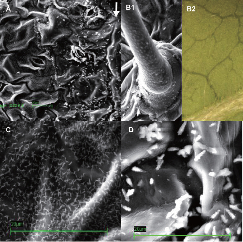
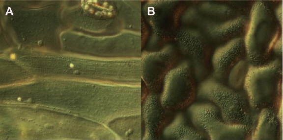
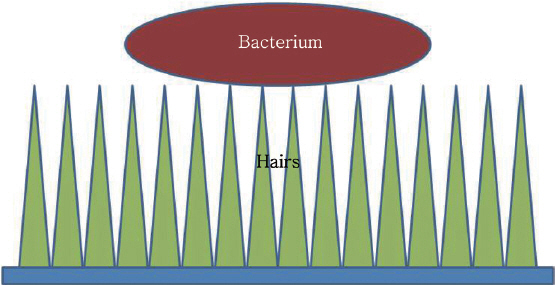
Ļ▓ĮņāüļČüļÅä ļåŹņŚģĻĖ░ņłĀņøÉ ņŗ£ĒŚśĒżņןņŚÉ ņ×¼ļ░░ņżæņØĖ Ēā£Ļ┤æ ĒÆłņóģņØś ņ×ÄņØä ņ▒äņĘ©ĒĢśņŚ¼ ņŻ╝ņé¼ņĀäņ×ÉĒśäļ»ĖĻ▓Į(VP1250, Zeiss) Ļ┤Ćņ░░ņŚÉ ņé¼ņÜ®ĒĢśņśĆļŗż. ņ▒äņĘ©ĒĢ£ ņ×ÄņØĆ 1├Ś107 cfu/mlņØś ļåŹļÅäļĪ£ X. axonopodis pv. glycinesļź╝ ĒؼņäØĒĢ£ ĒśäĒāüņĢĪņØä ļČäļ¼┤ĻĖ░ļĪ£ ņé┤ĒżĒĢśņŚ¼ ņāüņś©ņŚÉņä£ Ļ▒┤ņĪ░ĒĢ£ ļŗżņØī Ļ░ĆņÜ┤ļŹ░ ņŚĮļ¦źņØä ĒżĒĢ©ĒĢśņŚ¼ 5 mm ņĀĢļÅä Ēü¼ĻĖ░ņØś ņé¼Ļ░üĒśĢņ£╝ļĪ£ ņĀłļŗ©ĒĢśĻ│Ā ļÆĘ ļ®┤ņØ┤ ņ£äļĪ£ ņśżļÅäļĪØ ņŗ£ļŻīļīĆņŚÉ Ļ│ĀņĀĢņŗ£Ēé© ļŗżņØī ņāüņś©ņŚÉņä£ 3ņØ╝Ļ░ä Ļ▒┤ņĪ░ĒĢ£ Ēøä Ļ│©ļō£ņĮöĒīģņØä ĒĢśņŚ¼ Ļ┤Ćņ░░ņŚÉ ņé¼ņÜ®ĒĢśņśĆļŗż. ņ×ÄņØś SEM Ļ┤Ćņ░░ņŗ£ ņŚĮļ¦źĻ│╝ ņĢĮ 40 ┬Ąm ņĀĢļÅä ļ¢©ņ¢┤ņ¦ä ļČĆļČäļČĆĒä░ ņ×æņØĆ ņŚĮļ¬©Ļ░Ć ņ┤śņ┤śĒ׳ ļ░░ņŚ┤ļÉśņ¢┤ ņ׳ļŖö Ļ▓āņØ┤ Ļ┤Ćņ░░ļÉśņŚłņ£╝ļéś ņŚĮļ¦ź ņŻ╝ļ│ĆņØĆ ņØ┤ļ¤¼ĒĢ£ ņŚĮļ¬©Ļ░Ć Ļ┤Ćņ░░ļÉśņ¦Ć ņĢŖņĢśļŗż. ļśÉĒĢ£ ņŚĮļ¬©Ļ░Ć ņ׳ļŖö Ēæ£ļ®┤ņØĆ ļ©╝ņ¦Ćļéś ļ»ĖņāØļ¼╝ļō▒ ņØ┤ļ¼╝ņ¦łņØ┤ ļČĆņ░®ļÉśņ¢┤ ņ׳ļŖö Ļ▓āņØ┤ Ļ▒░ņØś Ļ┤Ćņ░░ļÉśņ¦Ć ņĢŖņØĆ ļ░śļ®┤ ņŚĮļ¦ź ņŻ╝ļ│ĆņØś ņŚĮļ¬©Ļ░Ć ņŚåļŖö Ēæ£ļ®┤ņØĆ ļ»ĖņāØļ¼╝ņØä ĒżĒĢ©ĒĢ£ ņØ┤ļ¼╝ņ¦łņØ┤ ļ¦ÄņØ┤ ļČĆņ░®ļÉśņ¢┤ ņ׳ļŖö Ļ▓āņØ┤ Ļ┤Ćņ░░ļÉśņŚłļŗż(Fig. 2A). TrichomņØä ņŗżņ▓┤Ēśäļ»ĖĻ▓Į Ļ┤Ćņ░░ņŗ£ ņ×Ä ņĀäņ▓┤ņŚÉ ļČäĒżĒĢśĻ│Ā ņ׳ņŚłņ£╝ļéś ņŻ╝ļĪ£ ņŚĮļ¦źņØä ļö░ļØ╝ ĒśĢņä▒ļÉśņ¢┤ ņ׳ņŚłļŗż(Fig. 2B2). Trichome ņØĆ ņØ╝ļ░śņĀüņ£╝ļĪ£ ņäĖĻĘĀņØ┤ ļČĆņ░®ĒĢśĻ▒░ļéś ņä£ņŗØĒĢśļŖö ņןņåīļź╝ ņĀ£Ļ│ĄĒĢśņŚ¼ ļ░£ļ│æņØä ņØ╝ņ£╝ĒéżļŖö ņøÉņØĖņØ┤ ļÉśļŖö Ļ▓āņ£╝ļĪ£ ņĢīļĀżņĀĖ ņ׳ņ£╝ļ»ĆļĪ£(Gets ļō▒, 1983; SchneiderņÖĆ Grogan, 1977), ņĮ® ņ×ÄņŚÉņä£ trichomeņØś ļČäĒżļź╝ Ļ│ĀļĀżĒĢĀ ļĢī trichome ļČĆņ£ä ņäĖĻĘĀ ļČĆņ░®ņØ┤ ņŚĮļ¦źņØä ļö░ļØ╝ ļ░£ļ│æņØ┤ ļÉśļŖö ņøÉņØĖ ņżæņØś ĒĢśļéśņØ╝ Ļ░ĆļŖźņä▒ņØĆ ņ׳ļŗż. ĻĘĖļ¤¼ļéś SEM Ļ┤Ćņ░░ņŗ£ trichomeņŚÉ ņäĖĻĘĀņØ┤ ļČĆņ░®ļÉ£ ļ¬©ņŖĄņØĆ Ļ┤Ćņ░░ĒĢĀ ņłś ņŚåņ¢┤ ņ¦üņĀæņĀüņØĖ ņāüĻ┤ĆĻ┤ĆĻ│äļŖö ņ░Šņ¦Ć ļ¬╗ĒĢśņśĆļŗż(Fig. 2B1). SEMņŚÉņä£ Ļ┤Ćņ░░ļÉ£ ņŚĮļ¬©ņØś Ēü¼ĻĖ░ļŖö 1 ┬Ąm ņØ┤ĒĢśļĪ£ ņØ╝ļ░śņĀüņØĖ ņäĖĻĘĀņØś Ēü¼ĻĖ░ļ│┤ļŗż ņ×æņĢśņ£╝ļ®░ ļÅÖņØ╝ĒĢ£ ņŗ£ļŻīņŚÉņä£ Ļ┤Ćņ░░ļÉ£ ņäĖĻĘĀņ£╝ļĪ£ ņČöņĀĢļÉśļŖö ļČĆņ░®ņ▓┤ļ│┤ļŗż ņ×æņØĆ Ļ▓āņØä ĒÖĢņØĖĒĢĀ ņłś ņ׳ņŚłļŗż(Fig. 2C, 2D). ņĮ®ņ×ÄņØś Ēæ£ļ®┤ ņäĖĒżļź╝ Ļ┤æĒĢÖĒśäļ»ĖĻ▓Įņ£╝ļĪ£ Ļ┤Ćņ░░ĒĢśĻĖ░ ņ£äĒĢ┤ ļ»ĖņäĖĒĢĆņģŗņ£╝ļĪ£ ņŚĮļ¦źļČĆņ£äĻ░Ć ĒżĒĢ©ļÉśĻ▓ī Ļ╗Źņ¦łņØä ļ▓ŚĻ▓© Ļ┤Ćņ░░ņŚÉ ņØ┤ņÜ®ĒĢśņśĆļŗż. Ļ┤æĒĢÖĒśäļ»ĖĻ▓Į Ļ┤Ćņ░░ņŗ£ 400ļ░░ ļ░░ņ£©ņŚÉņä£ļŖö ĒŖ╣ļ│äĒĢ£ ĻĄ¼ņĪ░ņĀü ņ░©ņØ┤ļŖö Ļ┤Ćņ░░ļÉśņ¦Ć ņĢŖņĢśņ£╝ļ®░ 1,000ļ░░ņØś ļ░░ņ£©ļĪ£ Ļ┤Ćņ░░ĒĢśņśĆņØä ļĢī ņŚĮļ¦źņŻ╝ņ£ä ņäĖĒżņŚÉņä£ļŖö ĒŖ╣ļ│äĒĢ£ ĻĄ¼ņĪ░Ļ░Ć Ļ┤Ćņ░░ļÉśņ¦Ć ņĢŖņĢśņ£╝ļéś(Fig. 3A) ņŚĮļ¦źĻ│╝ ļ¢©ņ¢┤ņ¦ä ļČĆņ£äņØś ņäĖĒżņŚÉņä£ļŖö ņŚĮļ¬©ļĪ£ ļ│┤ņØ┤ļŖö ņåīĒśĢņ×ģņ×ÉļōżņØ┤ ļŗżņłś Ļ┤Ćņ░░ļÉśņŚłļŗż(Fig. 3B). ņØ┤ļŖö SEM Ļ┤Ćņ░░Ļ│╝ ļÅÖņØ╝ĒĢśĻ▓ī ņŚĮļ¦ź ļČĆĻĘ╝ņŚÉņä£ļŖö Ļ┤Ćņ░░ļÉśņ¦Ć ņĢŖņĢśņ£╝ļ»ĆļĪ£ ņŚĮļ¬©ļĪ£ ĒīÉļŗ©ĒĢĀ ņłś ņ׳ņŚłļŗż. ļö░ļØ╝ņä£ ņŚĮļ¬©ļŖö Ļ┤æĒĢÖĒśäļ»ĖĻ▓Į 1,000ļ░░ņØś ļ░░ņ£©ņØ┤ļéś ņĀäņ×ÉĒśäļ»ĖĻ▓ĮņØä ņØ┤ņÜ®ĒĢĀ ļĢī Ļ┤Ćņ░░ņØ┤ Ļ░ĆļŖźĒĢ£ ļ»ĖņäĖņĪ░ņ¦üņ£╝ļĪ£ trichomeĻ│╝ ļŖö ĻĄ¼ļ│äļÉśļŖö ĻĄ¼ņĪ░ņ×äņØä ĒÖĢņØĖĒĢĀ ņłśĻ░Ć ņ׳ņŚłļŗż. ņŚĮļ¬©Ļ░Ć ņŚåļŖö ļČĆļČäņ£╝ļĪ£ ļ░£ļ│æņØ┤ ļÉśļŖö ņøÉņØĖņØĆ ņäĖĻĘĀņØ┤ Ēæ£ļ®┤ņŚÉ ļÅäļŗ¼ĒĢĀ Ļ▓ĮņÜ░ ņŚĮļ¬©Ļ░Ć ņØ╝ļ░śņĀüņØĖ ņäĖĻĘĀņØś Ēü¼ĻĖ░ļ│┤ļŗż ņ×æņĢäņä£ ņŗØļ¼╝ņ▓┤ ņ×Ä Ēæ£ļ®┤ņŚÉ ļČĆņ░®ĒĢśņ¦Ć ļ¬╗ĒĢśĻ│Ā ņŚĮļ¬©ņŚÉ ļČĆņ░®ĒĢśĻ▓ī ļÉśļŖöļŹ░ ņŚĮļ¬©ņØś Ēü¼ĻĖ░Ļ░Ć ņäĖĻĘĀļ│┤ļŗż ņ×æņ£╝ļ»ĆļĪ£ ņäĖĻĘĀĻ│╝ ņŗØļ¼╝ņ▓┤ņÖĆņØś ņĀæņ┤ēļ®┤ņØ┤ ņżäņ¢┤ļōżĻ▓ī ļÉśņ¢┤ ņäĖĻĘĀņØ┤ Ļ▓¼Ļ│ĀĒĢśĻ▓ī ļČĆņ░®ĒĢśņ¦Ć ļ¬╗ĒĢśĻ│Ā ņēĮĻ▓ī ļ¢©ņ¢┤ņĀĖ ļéśĻ░ĆļŖö Ļ▓āņ£╝ļĪ£ ņČöņĖĪļÉ£ļŗż(Fig. 4).
Fig.┬Ā2
Photograph of electron microscope of soybean leaf inoculated with Xanthomonas axonopodis pv. glycines. A: Bacteria and small hairs were observed only near vein, An arrow indicates vein, B1: Trichome, bacteria were not observed on the surface, B2: trichomes were observed over leaf, but mainly distributed on vein, C: small hairs were abundantly observed on the surface, D: bacteria-like-organism were attached near the vein.
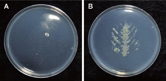
ņ×Ä Ēæ£ļ®┤ ļ»ĖņāØļ¼╝ņØś ļ░░ņ¢æ
ņäĖĻĘĀņØ┤ ņŚĮļ¦źņŻ╝ņ£äļĪ£ ļČĆņ░®ņØ┤ ļÉ£ļŗżļ®┤ ņŚĮļ¦ź ņŻ╝ņ£äņŚÉņä£ ņäĖĻĘĀņØ┤ Ļ▓ĆņČ£ļÉĀ Ļ▓āņØ┤ļ»ĆļĪ£ ņØ┤ļź╝ ņĢīņĢäļ│┤ĻĖ░ ņ£äĒĢ┤ Ēæ£ļ®┤ ļ»ĖņāØļ¼╝ņØś ļ░░ņ¢æņØä ņŗżņŗ£ĒĢśņśĆļŗż. ņś©ņŗżņŚÉņä£ ņ¦Ćļ”ä 12 cmņØś ĒżĒŖĖņŚÉ ņĮ®ņØä ĒīīņóģĒĢśņŚ¼ 5ļ¦łļööĻ╣īņ¦Ć ņ×Éļ×ÉņØä ļĢī PDA ļ░░ņ¦ĆņŚÉ 3ņØ╝Ļ░ä ļ░░ņ¢æĒĢ£ X. axonopodis pv. glycinesļź╝ ņé┤ĻĘĀļÉ£ ļŗłļōżļĪ£ ņ▒äņĘ©ĒĢśņŚ¼ ņé┤ĻĘĀņłśņŚÉ 1├Ś107 cfu/mlņØś ļåŹļÅäļĪ£ ĒؼņäØĒĢśņśĆļŗż. ĒؼņäØņĢĪņØĆ Ļ░ĆņĀĢņÜ® 500 ml ļČäļ¼┤ĻĖ░ļź╝ ņØ┤ņÜ®ĒĢśņŚ¼ ņżĆļ╣äĒĢ£ ņĮ® ņŗØļ¼╝ņ▓┤ņŚÉ ņé┤ĒżĒĢśņśĆņ£╝ļ®░ ļīĆņĪ░ĻĄ¼ļĪ£ļŖö ņé┤ĻĘĀņłśļź╝ ļČäļ¼┤ĒĢśņśĆļŗż. ņé┤Ēż 1ņØ╝ Ēøä ņ×ÄņØä ņ▒äņĘ©ĒĢśņŚ¼ potato dextrose agar(PDA) ļ░░ņ¦ĆņŚÉ ņ×Ä ļÆĘļ®┤ņØ┤ ņĀæņ┤ēļÉśĻ▓ī ņś¼ļ”░ ļŗżņØī ļ¼Ėņ¦łļ¤¼ņżīņ£╝ļĪ£ņŹ© Ēæ£ļ®┤ņØś ļ»ĖņāØļ¼╝ņØ┤ ļ░░ņ¦ĆņŚÉ ļČĆņ░®ļÉśļÅäļĪØ ĒĢ£ ļŗżņØī 25┬░C ļ░░ņ¢æĻĖ░ņŚÉņä£ 2ņØ╝Ļ░ä ļ░░ņ¢æĒĢśĻ│Ā Ļ▓░Ļ│╝ļź╝ Ļ┤Ćņ░░ĒĢśņśĆļŗż. ņé┤ĻĘĀņłśļź╝ ļČäļ¼┤ĒĢ£ ņ▓śļ”¼ņØś Ļ▓ĮņÜ░ ņäĖĻĘĀņØĆ ņĀäĒśĆ Ļ┤Ćņ░░ļÉśņ¦Ć ņĢŖņĢśņ£╝ļéś ļČłļ¦łļ”äļ│æĻĘĀņØä ņé┤ĒżĒĢ£ ņ▓śļ”¼ņØś Ļ▓ĮņÜ░ Ēæ£ļ®┤ļ░░ņ¢æņØä ĒĢ£ ņĮ® ņ×ÄņØś ņŚĮļ¦ź ņ£äņ╣śņŚÉņä£ ņäĖĻĘĀņØ┤ ņ×Éļ×Éņ£╝ļ®░(Fig. 5), ņØ┤ļź╝ ņ▒äņĘ©ĒĢśņŚ¼ Lee ļō▒(2013)ņØś ļ░®ļ▓ĢņŚÉ ļö░ļØ╝ PCRņØä ņŗżņŗ£ĒĢśņśĆņØä ļĢī ļČłļ¦łļ”äļ│æĻĘĀņ×äņØä ĒÖĢņØĖĒĢĀ ņłś ņ׳ņŚłļŗż(Data not shown). ņØ┤ļ¤¼ĒĢ£ Ļ▓░Ļ│╝ļŖö SEMņŚÉņä£ Ļ┤Ćņ░░ĒĢ£ ņŚĮļ¬©Ļ░Ć ņŚåļŖö ņ£äņ╣śņŚÉ ņäĖĻĘĀņØ┤ ļČĆņ░®ļÉśņ¢┤ ņ׳ļŖö Ļ▓░Ļ│╝ņÖĆ ļÅÖņØ╝ĒĢśļŗż ĒĢĀ ņłś ņ׳ļŗż. ļśÉĒĢ£, ņŚĮļ¦źņŚÉ ļŗżņłś ņĪ┤ņ×¼ĒĢśļŖö trichomeņØ┤ ņŚĮļ¦źņØä ļö░ļØ╝ ļ│æņ¦ĢņØ┤ ņāØĻĖ░ļŖö ņøÉņØĖņżæņØś ĒĢśļéśņØ╝ Ļ░ĆļŖźņä▒ļÅä ņ׳ņ£╝ļéś ņØ┤ Ļ▓ĮņÜ░ trichomeņØ┤ ņŚĮļ¦źņŚÉ ļ¦ÄņØ┤ ļČäĒżĒĢśņ¦Ćļ¦ī ņ×Ä ņĀäņ▓┤ņŚÉ ņé░ņ×¼ĒĢ┤ ņ׳ĻĖ░ ļĢīļ¼ĖņŚÉ ņäĖĻĘĀ ļ░░ņ¢æņŗ£ ņ×Ä ņĀäļ®┤ņŚÉ ņäĖĻĘĀņØ┤ ļ░░ņ¢æņØ┤ ļÉśņ¢┤ņĢ╝ ĒĢĀ Ļ▓āņØ┤ļŗż. ĻĘĖļ¤¼ļ»ĆļĪ£ ļČłļ¦łļ”äļ│æņØś ļ│æņ¦ĢņØ┤ ņŚĮļ¦ź ņŻ╝ļ│Ćņ£╝ļĪ£ ĒśĢņä▒ļÉśļŖö ņøÉņØĖņØĆ ņŚĮļ¬©Ļ░Ć ļ¼╝ļ”¼ņĀü ņןļ▓Į ņŚŁĒĢĀņØä ĒĢśņŚ¼ ņŚĮļ¬©Ļ░Ć ņĪ┤ņ×¼ĒĢśļŖö ļČĆņ£äļŖö ļ│æņøÉĻĘĀņØś ļČĆņ░®ņØ┤ ņĀĆĒĢ┤ļÉ©ņ£╝ļĪ£ ņŚĮļ¬©Ļ░Ć ņŚåļŖö ņŚĮļ¦ź ņŻ╝ņ£äļĪ£ ļ│æņøÉĻĘĀņØ┤ ļČĆņ░®ļÉśņ¢┤ ļ░£ļ│æņØ┤ ļÉśļŖö Ļ▓āņ£╝ļĪ£ ĒīÉļŗ©ĒĢĀ ņłś ņ׳ļŗż.
 PDF Links
 PubReader
 Full text via DOI
 Download Citation
 Print
Print


