


ņä£ ļĪĀ
ĻĄŁĒÖöļŖö ĻĄŁĒÖöĻ│╝ņŚÉ ņåŹĒĢśļŖö ņŚ¼ļ¤¼ĒĢ┤ņé┤ņØ┤ ņŗØļ¼╝ļĪ£ Ļ┤ĆņāüņÜ®, ņĢĮņÜ®Ļ│╝ ņŗØņÜ®ņ£╝ļĪ£ ĒÖ£ņÜ®ļÉ£ļŗż. ĻĄŁĒÖöļŖö ņŖżĒāĀļŗżļō£ ĻĄŁĒÖö(ļīĆĻĄŁ)Ļ│╝ ņŖżĒöäļĀłņØ┤ ĻĄŁĒÖö(ņåīĻĄŁ)ņ£╝ļĪ£ ĻĄ¼ļČäĒĢĀ ņłś ņ׳ņ£╝ļ®░, ĻĄŁļé┤ņŚÉņä£ļŖö ņŖżĒāĀļŗżļō£ ĻĄŁĒÖöņØś ņ×¼ļ░░ļ®┤ņĀü(242.9 ha)ņØ┤ ņĀäņ▓┤ ĻĄŁĒÖö ņ×¼ļ░░ļ®┤ņĀü(309.1 ha)ņØś 78.6% ļź╝ ņ░©ņ¦ĆĒĢ£ļŗż(Ministry of Agriculture, Food and Rural Affairs, 2020). ĻĄŁļé┤ ņ×¼ļ░░ņ¦Ć Ļ░ĆņÜ┤ļŹ░ Ļ▓Įļé© ņ░ĮņøÉ ļ░Å ļČĆņé░ ņ¦ĆņŚŁņØĆ ņŗĀļ¦ł, ļ░▒ļ¦ł, ļ░▒ņäĀ ĒÆłņóģ ļō▒ņØä ņŖżĒāĀļŗżļō£ ĻĄŁĒÖöļź╝ ņŗ£ņäżņŚÉņä£ ņØ╝ļ│Ė ņłśņČ£ņÜ®ņ£╝ļĪ£ ņ×¼ļ░░ĒĢśĻ│Ā ņ׳ņ£╝ļ®░ ĻĘĖ ņ×¼ļ░░ļ®┤ņĀüņØĆ 125.5 haņŚÉ ļŗ¼ĒĢ£ļŗż(Ministry of Agriculture, Food and Rural Affairs, 2020).
ĻĄŁĒÖö ņ×¼ļ░░ņŚÉņä£ ņŻ╝ļĪ£ ļ¼ĖņĀ£Ļ░Ć ļÉśļŖö Orthotospovirus ņåŹņŚÉ ņåŹĒĢśļŖö ļ░öņØ┤ļ¤¼ņŖżļōżņØĆ ĒåĀļ¦łĒåĀļ░śņĀÉņ£äņĪ░ļ░öņØ┤ļ¤¼ņŖż(Tomato spotted wilt virus, TSWV)ņÖĆ ĻĄŁĒÖöņżäĻĖ░Ļ┤┤ņĀĆļ░öņØ┤ļ¤¼ņŖż(Chrysanthemum stem necrosis virus, CSNV)ļĪ£ ņĢīļĀżņĀĖ ņ׳ļŗż(Chung ļō▒, 2006; Yoon ļō▒, 2017). TSWVņÖĆ CSNVļŖö 3Ļ░£ņØś (ŌĆÆ) stranded Ēś╣ņØĆ ambisense RNAņØś ļČäņĀł Ļ▓īļåł(L, M ļ░Å S segments) ĒśĢĒā£ļĪ£ ņØ┤ļŻ©ņ¢┤ņĀĖ ņ׳ņ£╝ļ®░, ņŗØļ¼╝Ļ│╝ ņŗØļ¼╝ ņé¼ņØ┤ņØś ņĀäņŚ╝ņØĆ ņŻ╝ļĪ£ ņ┤Øņ▒äļ▓īļĀłņŚÉ ņØśĒĢ┤ ļ¦żĻ░£ļÉśņ¢┤ ĻĄŁĒÖö, Ļ│ĀņČö, ĒåĀļ¦łĒåĀ, ņ×Īņ┤łļźś ļō▒ 1,090ņŚ¼ņóģ ņŗØļ¼╝ņØä Ļ░ÉņŚ╝ņŗ£Ēé©ļŗż(Parrella ļō▒, 2003). ĻĄŁļé┤ņŚÉņä£ TSWVļŖö 2003ļģä ņśłņé░ņ¦ĆņŚŁ ĒīīĒöäļ”¼ņ╣┤ ņ×¼ļ░░ļåŹĻ░ĆņŚÉņä£ ņ▓śņØī ļ░£Ļ▓¼ļÉ£ ņØ┤Ēøä, ļŗżņ¢æĒĢ£ ņøÉņśłņ×æļ¼╝ņŚÉņä£ Ēö╝ĒĢ┤Ļ░Ć ĒÖĢņØĖļÉśņŚłņ£╝ļ®░ ĻĄŁĒÖöņŚÉņä£ļŖö 2006ļģä ņČ®ļé© Ēā£ņĢł ļ░Å ņśłņé░ņØś ĻĄŁĒÖö ņŗ£ņäżĒĢśņÜ░ņŖż(ĒÆłņóģ: ņŗĀļ¦ł, ĒÖöļ×æ)ņŚÉņä£ TSWVĻ░Ć ņ▓śņØī ļ│┤Ļ│ĀļÉśņŚłļŗż(Chung ļō▒, 2006; Kim ļō▒, 2004).
ņĀä ņäĖĻ│äņĀüņ£╝ļĪ£ ĻĄŁĒÖö ņ×¼ļ░░ņŚÉņä£ ļ¼ĖņĀ£ļÉśļŖö ĒĢ┤ņČ®ņØĆ ņ┤Øņ▒äļ▓īļĀł, ņ¦äļö¦ļ¼╝, ņØæņĢĀ ļō▒ņØ┤ļ®░ ĻĘĖ ņżæ Ļ░Ćņן Ēö╝ĒĢ┤ļź╝ ņŻ╝ļŖö ĒĢ┤ņČ®ņØĆ ņ┤Øņ▒äļ▓īļĀłļĪ£ ļ│┤Ļ│ĀļÉśņŚłļŗż(Lewis, 1997). ņ┤Øņ▒äļ▓īļĀłļŖö ņäĖĻ│äņĀüņ£╝ļĪ£ ņĢĮ 5,500ņŚ¼ņóģņØ┤ ļ│┤Ļ│ĀļÉśņŚłņ£╝ļ®░ 87ņóģņØ┤ Ļ▓ĮņĀ£ņĀüņ£╝ļĪ£ ņżæņÜöĒĢ£ ņ×æļ¼╝ņŚÉ Ēö╝ĒĢ┤ļź╝ ņŻ╝Ļ│Ā ņ׳ļŖö Ļ▓āņ£╝ļĪ£ ļ│┤Ļ│ĀļÉśņŚłļŗż(Demirozer ļō▒, 2012). ĻĄŁļé┤ņŚÉņä£ļŖö ĻĄŁĒÖö, Ļ│ĀņČö, Ēīī, ņśżņØ┤ ļō▒ņŚÉņä£ Ļ▓ĮņĀ£ņĀüņ£╝ļĪ£ Ēü░ Ēö╝ĒĢ┤ļź╝ ņŻ╝ļŖö ņ┤Øņ▒äļ▓īļĀł ņóģļźśļĪ£ļŖö Ēīīņ┤Øņ▒äļ▓īļĀł(Thrips tabaci), ĻĮāļģĖļ×æņ┤Øņ▒äļ▓īļĀł(Frankliniella occidentalis), ļ│╝ļĪØņ┤Øņ▒äļ▓īļĀł(Scirtothrips dorsalis) ļ░Å ņśżņØ┤ņ┤Øņ▒äļ▓īļĀł(T. palmi)ļĪ£ ļ│┤Ļ│ĀļÉśņŚłņ£╝ļ®░, ņĢ×ņä£ ĻĖ░ņłĀļÉ£ ņ┤Øņ▒äļ▓īļĀł ņóģņØä ĒżĒĢ©ĒĢśņŚ¼ TSWVļź╝ ļ¦żĻ░£ĒĢśļŖö ņ┤Øņ▒äļ▓īļĀłļŖö 11ņóģņØ┤ ļ│┤Ļ│ĀļÉśņŚłļŗż(Hyun ļō▒, 2012; MorseņÖĆ Hoddle, 2006). ņØ┤ļōż ņ┤Øņ▒äļ▓īļĀłļźśļŖö ņé┤ņČ®ņĀ£ņŚÉ Ļ░ĢĒĢ£ ņĢĮņĀ£ ņĀĆĒĢŁņä▒ņØä Ļ░Ćņ¦Ćļ®░, ņ×Ä ļÆĘļ®┤ ļśÉļŖö ĻĮā ņåŹņŚÉ ņł©ļŖö ĒŖ╣ņä▒ ļĢīļ¼ĖņŚÉ ņĢĮņĀ£ ļ░®ņĀ£Ļ░Ć ņ¢┤ļĀżņÜ┤ ĒĢ┤ņČ®ņ£╝ļĪ£ ņĢīļĀżņĀĖ ņ׳ļŗż(Han ļō▒, 1998; Lee, 1996; SchmidtņÖĆ Frey, 1995). ņä▒ņČ®ņØĆ ĒÖ®ņāē ļśÉļŖö ĒÖ®Ļ░łņāēņØä ļØäļ®░ ĒÅēĻĘĀ Ēü¼ĻĖ░ļŖö 1.17 mm ļé┤ņÖĖņØ┤ļ®░ ņŗØļ¼╝ņ▓┤ ņĪ░ņ¦ü ņåŹņŚÉ ņé░ļ×ĆĒĢśļŖö Ļ▓āņ£╝ļĪ£ ņĢīļĀżņĀĖ ņ׳ļŗż. ņ×ÄņŚÉņä£ ļČĆĒÖöĒĢ£ ņĢīņŚÉņä£ 1ļĀ╣ ņĢĮņČ® ļŗ©Ļ│äļź╝ Ļ▒░ņ│É 2ļĀ╣ ņĢĮņČ® ņäĖļīĆļź╝ Ļ▒░ņ╣śļ®░ ņØ┤Ēøä ņŻ╝ļĪ£ ĒåĀņ¢æņ£╝ļĪ£ ņØ┤ļÅÖĒĢśņŚ¼ ĒåĀņ¢æņŚÉņä£ ļ▓łļŹ░ĻĖ░ Ļ│╝ņĀĢ(ņĀäņØśņÜ®, ĒøäņØśņÜ®)ņØä Ļ▒░ņ╣£ ļŗżņØī ņä▒ņČ®ņ£╝ļĪ£ ņÜ░ĒÖö Ēøä ņ¦ĆņāüļČĆļĪ£ ņØ┤ļÅÖĒĢśņŚ¼ ņŗØļ¼╝ņØä Ļ░ĆĒĢ┤ĒĢ£ļŗż. ĻĄŁĒÖöņŚÉņä£ļŖö ļ¼ĖņĀ£ļÉśļŖö ņ┤Øņ▒äļ▓īļĀłļźśļŖö ĻĮāļģĖļ×æņ┤Øņ▒äļ▓īļĀłņÖĆ ļīĆļ¦īņ┤Øņ▒äļ▓īļĀł(F. intonsa)ņ£╝ļĪ£ ņĢīļĀżņĀĖņ׳ņ£╝ļéś(OkazakiņÖĆ Sakurai, 2005; Okuda ļō▒ 2013), ļģĖņ¦Ćņ×¼ļ░░ņŚÉ ļ╣äĒĢ┤ ņŗ£ņäżņ×¼ļ░░ ĻĄŁĒÖöņŚÉņä£ ĻĮāļģĖļ×æņ┤Øņ▒äļ▓īļĀłĻ░Ć ļåÆņØĆ ļ░ĆļÅäļĪ£ ļ░£ņāØļÉśĻ│Ā ņ׳ļŗż(Park ļō▒, 2002; Yoon ļō▒, 2020). ņ┤Øņ▒äļ▓īļĀłļŖö ņŻ╝ļĪ£ ņ¢┤ļ”░ ĻĄŁĒÖö ņ×ÄņØä Ļ░ĆĒĢ┤ĒĢśņŚ¼ ņ×ÄņØś ĻĖ░ĒśĢņØä ņ┤łļלĒĢśĻ│Ā, ĻĄŁĒÖö ĻĮāņØ┤ Ēö╝ĻĖ░ ņŗ£ņ×æĒĢśļ®┤ ņ£ĀņČ®Ļ│╝ ņä▒ņČ®ļōżņØ┤ ĻĮā ņĢłņ£╝ļĪ£ ļōżņ¢┤Ļ░Ć ņ¢┤ļ”░ ĻĮāņ×ÄņØä Ļ░ēņĢä ņāüņ▓śļź╝ ļéĖļŗż. ņØ┤Ēøä ĻĮāņ×ÄņØ┤ ņĀäĻ░£ļÉśļ®┤ ņāüņ▓śļČĆņ£äĻ░Ć ņŗ£ļōżĻ│Ā ĒāłņāēļÉśņ¢┤ ĻĄŁĒÖöņØś ņāüĒÆł Ļ░Ćņ╣śļź╝ ļ¢©ņ¢┤ļ£©ļ”¼Ļ▓ī ļÉ£ļŗż. ņØ┤ļ¤¼ĒĢ£ ņ┤Øņ▒äļ▓īļĀłļŖö TSWV ļ░Å CSNVļź╝ ļ¦żĻ░£ĒĢ£ļŗż. ņ┤Øņ▒äļ▓īļĀłļŖö 1ŌĆÆ2ļĀ╣ ņĢĮņČ® ņŗ£ĻĖ░ņŚÉ TSWVļź╝ ĒØĪņ”ÖĒĢśņŚ¼ ņśüņåŹņĀäņŚ╝(persistent transmission)ņ£╝ļĪ£ ņĀäņŚ╝, ĒÖĢņé░ņŗ£ĒéżļŖö Ļ▓āņ£╝ļĪ£ ņĢīļĀżņĀĖ ņ׳ņ£╝ļéś ļŗżļźĖ ņ┤Øņ▒äļ▓īļĀł ņóģĻ│╝ ļŗżļź┤Ļ▓ī ĻĮāļģĖļ×æņ┤Øņ▒äļ▓īļĀłņÖĆ ļīĆļ¦īņ┤Øņ▒äļ▓īļĀłļŖö ņä▒ņČ® ļŗ©Ļ│äņŚÉņä£ļÅä TSWVļź╝ ņĀäņŚ╝ņŗ£Ēé©ļŗż(Inoue ļō▒ 2004; Maris, 2004; Wijkamp ļō▒, 1996).
ņ┤Øņ▒äļ▓īļĀł ņóģ ļÅÖņĀĢļ▓Ģņ£╝ļĪ£ internal transcribed spacer 2 (ITS2) ņśüņŚŁņØś PCR ņé░ļ¼╝ņØä ņĀ£ĒĢ£ĒÜ©ņåī RsaIņ£╝ļĪ£ ņ▓śļ”¼ĒĢśļŖö polymerase chain reactionŌĆÆrestriction fragment length polymorphism ļ▓Ģ ļ░Å ITS2 ņ£ĀņĀäņ×ÉņØś ņóģ ĒŖ╣ņØ┤ņĀüņØĖ ĒöäļØ╝ņØ┤ļ©Ėļź╝ ņØ┤ņÜ®ĒĢśņŚ¼ PCR ļ░Å ņŚ╝ĻĖ░ņä£ņŚ┤ ļČäņäØņØä ņłśĒ¢ēĒĢśļŖö ļ░®ļ▓Ģ ļō▒ņØ┤ Ļ░£ļ░£ļÉśņ¢┤ ņ׳ļŗż(Brunner ļō▒, 2002; NakaharaņÖĆ Minoura, 2015; TodaņÖĆ Komazaki, 2002; Yeh ļō▒, 2014, 2015). ņØ╝ļ░śņĀüņ£╝ļĪ£ Ļ░ÉņŚ╝ļÉ£ ņŗØļ¼╝ņ▓┤ņŚÉņä£ TSWVļź╝ Ļ▓ĆņČ£ĒĢśĻĖ░ ņ£äĒĢ┤ ĒÜ©ņåīĻ▓░ĒĢ®ļ®┤ņŚŁĒØĪņ░®Ļ▓Ćņé¼ļ▓Ģ(enzyme-linked immunosorbent assay)ņÖĆ ņŚŁņĀäņé¼ņżæĒĢ®ĒÜ©ņåīņŚ░ņćäļ░śņØæļ▓Ģ(reverse transcriptionŌĆÆpolymerase chain reaction, RT-PCR), ņĀĢļ¤ēņĀü ņŚŁņĀäņé¼ņżæĒĢ®ĒÜ©ņåīņŚ░ņćäļ░śņØæļ▓Ģ(quantitative transcriptionŌĆÆpolymerase chain reaction)ņØ┤ ļ│┤Ļ│ĀļÉśņŚłļŗż(Chung ļō▒, 2006, Yoon ļō▒, 2017). ļśÉĒĢ£ ņ┤Øņ▒äļ▓īļĀłņŚÉņä£ TSWVļź╝ Ļ▓ĆņČ£ĒĢśĻĖ░ ņ£äĒĢśņŚ¼ ņĢ×ņä£ ĻĖ░ņłĀĒĢ£ 3Ļ░Ćņ¦Ć ļ░®ļ▓ĢļōżņØä ņØ┤ņÜ® ĻĖ░ņłĀņØ┤ ļ│┤Ļ│ĀļÉśņŚłņ£╝ļéś ņØ┤ļ¤¼ĒĢ£ ļ░®ļ▓ĢņØĆ ņŗ£Ļ░äņØ┤ ļ¦ÄņØ┤ ņåīņÜöļÉśĻ▒░ļéś ļ»╝Ļ░ÉļÅäĻ░Ć ļ¢©ņ¢┤ņ¦ĆĻ│Ā ĻĄ¼ņ×ģ ņןļ╣äĻ░Ć ļ╣äņŗĖļŗżļŖö ļŗ©ņĀÉņØ┤ ņ׳ņ£╝ļ®░ ļśÉĒĢ£ ņ┤Øņ▒äļ▓īļĀł ņóģ ļÅÖņĀĢņØä ļ│äļÅäļĪ£ ņłśĒ¢ēĒĢ┤ņĢ╝ ĒĢśļŖö ļ▓łĻ▒░ļĪ£ņøĆņØ┤ ņ׳ļŗż(Bandla ļō▒, 1994; Boonham ļō▒, 2002; Cho ļō▒, 1989; Tsuda ļō▒, 1994).
ņØ┤ļ▓ł ņŚ░ĻĄ¼ņŚÉņä£ TSWV ņÖĖĒö╝ļŗ©ļ░▒ņ¦ł(N) ņ£ĀņĀäņ×É ļ░Å ņ┤Øņ▒äļ▓īļĀł ITS2 ļČĆļČäņŚÉ ĒŖ╣ņØ┤ņĀüņØĖ ĒöäļØ╝ņØ┤ļ©Ėļź╝ ņØ┤ņÜ®ĒĢśņŚ¼ ņ┤Øņ▒äļ▓īļĀł 1ļ¦łļ”¼ņŚÉņä£ TSWV ļ│┤ļÅģ ņŚ¼ļČĆ ļ░Å ņ┤Øņ▒äļ▓īļĀłņØś ņóģ ļÅÖņĀĢņØä ļÅÖņŗ£ņŚÉ ĒÖĢņØĖĒĢĀ ņłś ņ׳ļŖö ņ¦äļŗ©ļ▓ĢņØä Ļ░£ļ░£ĒĢśņśĆļŗż. ņØ┤ļź╝ ņØ┤ņÜ®ĒĢśņŚ¼ ĻĄŁĒÖö ņ×¼ļ░░ ņś©ņŗżņŚÉņä£ ņłśņ¦æĒĢ£ ņ┤Øņ▒äļ▓īļĀłņŚÉņä£ TSWV ļ│┤ļÅģļźĀ ļ░Å ņ┤Øņ▒äļ▓īļĀł ņÜ░ņĀÉņóģņØä ļÅÖņŗ£ņŚÉ ņĪ░ņé¼Ļ░Ć Ļ░ĆļŖźĒĢ©ņØä ļ│┤ņŚ¼ņŻ╝ļ®░ ņ┤Øņ▒äļ▓īļĀłļź╝ ĒåĄĒĢ£ TSWVņØś ņŗ£ņäżļé┤ ņ£Āņ×ģņŗ£ĻĖ░ņÖĆ ļ░£ņāØ Ļ▓ĮļĪ£ ļō▒ TSWV ņŚŁĒĢÖ ņŚ░ĻĄ¼ņŚÉ ĒÖ£ņÜ®ĒĢĀ ņłś ņ׳ņØīņØä ņśłņŗ£ĒĢ┤ņżĆļŗż. ņØ┤ļ¤¼ĒĢ£ ņŚ░ĻĄ¼ļŖö ņŗ£ņäżĻĄŁĒÖöņŚÉņä£ TSWV ļ│æņ¦ĢņØ┤ ļéśĒāĆļéśĻĖ░ ņØ┤ņĀä ņ┤Øņ▒äļ▓īļĀłļź╝ ņØ┤ņÜ®ĒĢ£ TSWV ņśłņ░░ņŚÉ ĒÖ£ņÜ®ĒĢĀ ņłś ņ׳ņØä Ļ▓āņØ┤ļØ╝ ņāØĻ░üļÉ£ļŗż.
ņ×¼ļŻī ļ░Å ļ░®ļ▓Ģ
ļ░öņØ┤ļ¤¼ņŖżņøÉ ļ░Å ņ┤Øņ▒äļ▓īļĀł ņłśņ¦æ
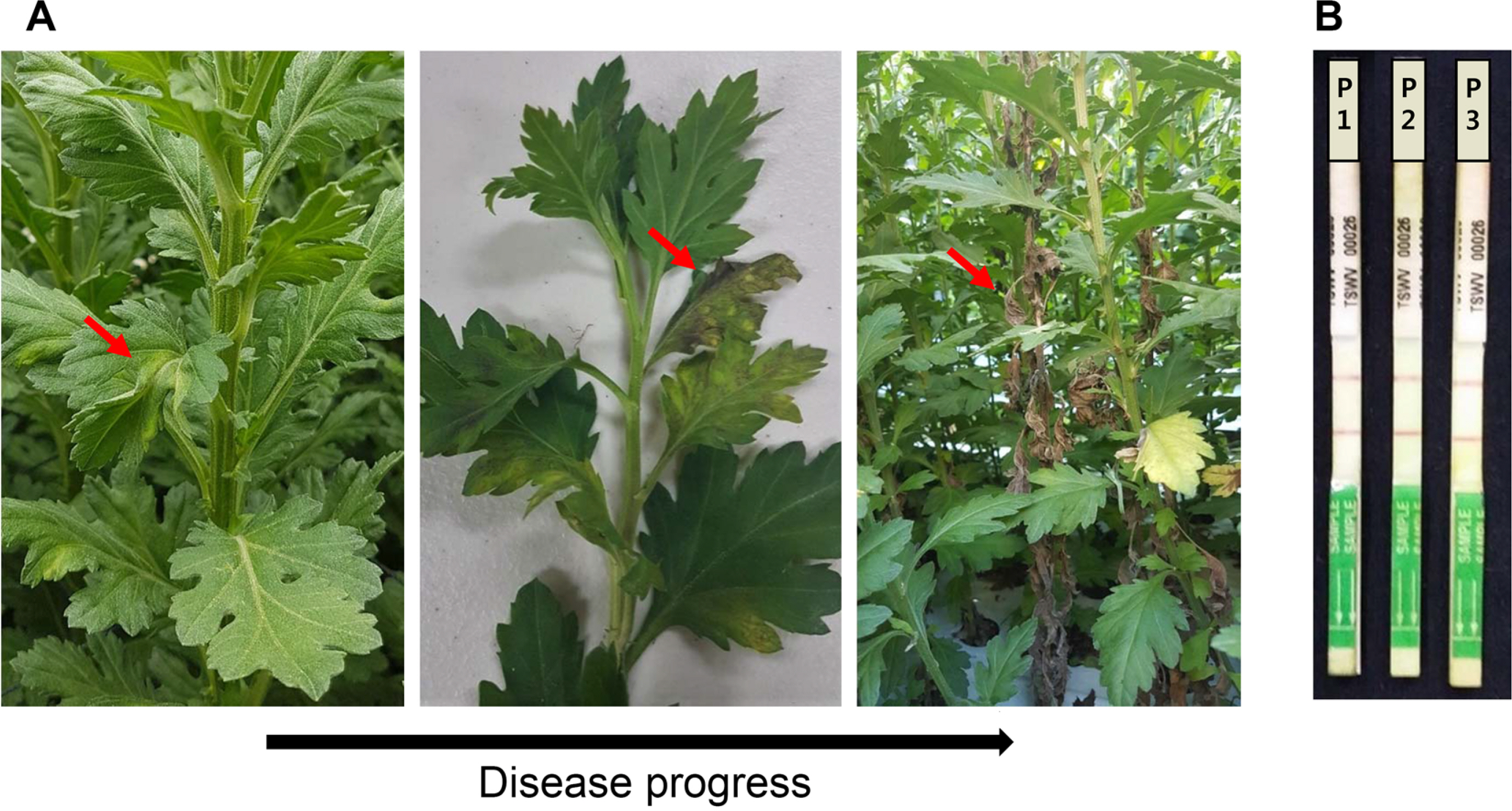
Ļ▓Įļé© ņ░ĮņøÉņŗ£ ĻĄŁĒÖöņ×¼ļ░░ ņŗ£ņäż ĒĢśņÜ░ņŖżņŚÉņä£ ņłśņ¦æĒĢ£ TSWV Ļ░ÉņŚ╝ ĻĄŁĒÖö(ļ░▒ļ¦ł ĒÆłņóģ)ļź╝ Ļ│Ąņŗ£ņ×¼ļŻīļĪ£ ņé¼ņÜ®ĒĢśņśĆļŗż. TSWV Ļ░ÉņŚ╝ ĻĄŁĒÖöļŖö ņāüņŚĮĻ│╝ ņżäĻĖ░Ļ░Ć ļÆżĒŗĆņ¢┤ņĀĖ ņ׳Ļ│Ā ļ│æņØ┤ ņ¦äņĀäļÉśļ®┤ ņ×ÄĻ│╝ ņżäĻĖ░ ļüØņŚÉ Ļ┤┤ņé¼ņ”ØņāüņØ┤ ļéśĒāĆļéś Ļ│Āņé¼ĒĢśņśĆļŗż(Fig. 1). TSWV Ļ░ÉņŚ╝ ņĪ░ņé¼ļŖö SEB1 ņÖäņČ®ņĢĪ(Agdia, Beltsville, MD, USA)ņŚÉ ņ£äņØś ĻĄŁĒÖöņØś ļ│æļōĀ ņ×ÄņØä ļ¦łņćäĒĢ£ ņĪ░ņ”ÖņĢĪņŚÉ TSWV ĒŖ╣ņØ┤ņĀü ImmunoStrip (Agdia)ņØä ļŗ┤ĻĘ╝ Ēøä ļéśĒāĆļéśļŖö Ļ▓░Ļ│╝ļĪ£ ĒÖĢņØĖĒĢśņśĆļŗż. TSWVĻ░Ć ņ¦äļŗ©ļÉ£ ĻĄŁĒÖö ņ×ÄņØä 0.1 M potassium phosphate (pH 7.2) ņÖäņČ®ņĢĪņŚÉņä£ ļ¦łņćäĒĢ£ ņĪ░ņ”ÖņĢĪņØä carborundum (Thermo Fisher Scientific, Waltham, MA, USA)ņØä ļ┐īļ”░ ņ▓Łņ¢æĻ│ĀņČö(Capsicum annuum L.)ņŚÉ ņĀæņóģĒĢśņśĆļŗż. 2ņŻ╝ Ēøä ņāüņŚĮņŚÉ ĻĮāļģĖļ×æņ┤Øņ▒äļ▓īļĀł ļ░Å ļīĆļ¦īņ┤Øņ▒äļ▓īļĀł 1ŌĆÆ2ļĀ╣ ņĢĮņČ®ņØä ņŗØļ¼╝ņ▓┤ļŗ╣ 30ļ¦łļ”¼ņö® ļČōņØä ņØ┤ņÜ®ĒĢśņŚ¼ ņś¼ļĀż TSWVļź╝ ļ│┤ļÅģņŗ£Ēé© ļŗżņØī 7ņØ╝ ņØ┤ņāü ņ£Āņ¦Ćņŗ£ņ╝£ ņä▒ņČ®ņ£╝ļĪ£ ņ£ĀļÅäļÉ£ ĻĮāļģĖļ×æņ┤Øņ▒äļ▓īļĀł ļśÉļŖö ļīĆļ¦īņ┤Øņ▒äļ▓īļĀłļź╝ ņØ┤Ēøä ņŗżĒŚśņŚÉ ņé¼ņÜ®ĒĢśņśĆļŗż.
Ļ░£ļ░£ĒĢ£ ĻĖ░ņłĀņØä ņ”Øļ¬ģĒĢśĻĖ░ ņ£äĒĢśņŚ¼ Ļ▓Įļé© ņ░ĮņøÉņŗ£ ļ░Å ņČ®ļé© Ēā£ņĢłņ¦ĆņŚŁ ĻĄŁĒÖö ņ×¼ļ░░ņ¦ĆņŚÉņä£ ņ┤Øņ▒äļ▓īļĀłļź╝ Ļ░ü 43, 102ļ¦łļ”¼ļź╝ Ēżņ¦æĒĢśņśĆņ£╝ļ®░ Ļ░ü ņ┤Øņ▒äļ▓īļĀłļź╝ ņŗżņ▓┤Ēśäļ»ĖĻ▓Į(Carl Zeiss, Jena, Germany)ņØä ņØ┤ņÜ®ĒĢśņŚ¼ Ļ░äņØ┤ ļÅÖņĀĢņØä ņłśĒ¢ēĒĢśņśĆĻ│Ā Ļ░£ņ▓┤ļ│äļĪ£ 70% ņŚÉĒāäņś¼ņØ┤ ļŗ┤ĻĖ┤ 1.5 ml Eppendorf tubeņŚÉ ļäŻņ¢┤ ŌłÆ20┬░CņŚÉ ļ│┤Ļ┤ĆĒĢśĻ▒░ļéś ĒĢĄņé░ ņČöņČ£ņŚÉ ņé¼ņÜ®ĒĢśņśĆļŗż.
ĒĢĄņé░ ņČöņČ£
ĒĢĄņé░ņČöņČ£ņØä ņ£äĒĢ┤ ņ”ØņāüņØ┤ ļ│┤ņØ┤ļŖö ĻĄŁĒÖö ņ×Ä 100 mg Ēś╣ņØĆ ņ┤Øņ▒äļ▓īļĀł 1ŌĆÆ10ļ¦łļ”¼(0.05ŌĆÆ0.5 mg)ļź╝ ņĢĪņ▓┤ņ¦łņåīĻ░Ć ļōżņ¢┤ ņ׳ļŖö 1.5 ml Eppendorf tubeņŚÉ ļäŻņØĆ ļŗżņØī ļ¦łņćäĒĢśņśĆļŗż. ņØ┤Ēøä ņĀ£ņĪ░ņé¼ņØś ņČöņ▓£ ļ░®ļ▓ĢļīĆļĪ£ Plant RNA/DNA purification kit (Norgenbiotek, Thorold, ON, Canada)ļź╝ ņØ┤ņÜ®ĒĢśņŚ¼ ĒĢĄņé░ņØä ņČöņČ£ĒĢśņśĆļŗż. ņČöņČ£ļÉ£ ĒĢĄņé░ņØś ņł£ļÅäļŖö NanoDrop ļČäĻ┤æĻ┤æļÅäĻ│ä(Thermo Fisher Scientific)ļź╝ ņØ┤ņÜ®ĒĢśņŚ¼ ĒØĪĻ┤æļÅä 260 nmņÖĆ 280 nm ļ╣äņ£© (A260/A280)ļĪ£ Ļ▓░ņĀĢĒĢśņśĆļŗż. ņČöņČ£ļÉ£ ĒĢĄņé░ņØĆ ņŗżĒŚśņŚÉ ļ░öļĪ£ ņé¼ņÜ®ĒĢśĻ▒░ļéś ŌĆÆ80┬░C ļāēļÅÖĻ│ĀņŚÉ ļ│┤Ļ┤ĆĒĢśņŚ¼ ņØ┤Ēøä ņŗżĒŚśņŚÉ ņé¼ņÜ®ĒĢśņśĆļŗż.
ĒöäļØ╝ņØ┤ļ©Ė ņĀ£ņ×æ ļ░Å RT-PCR
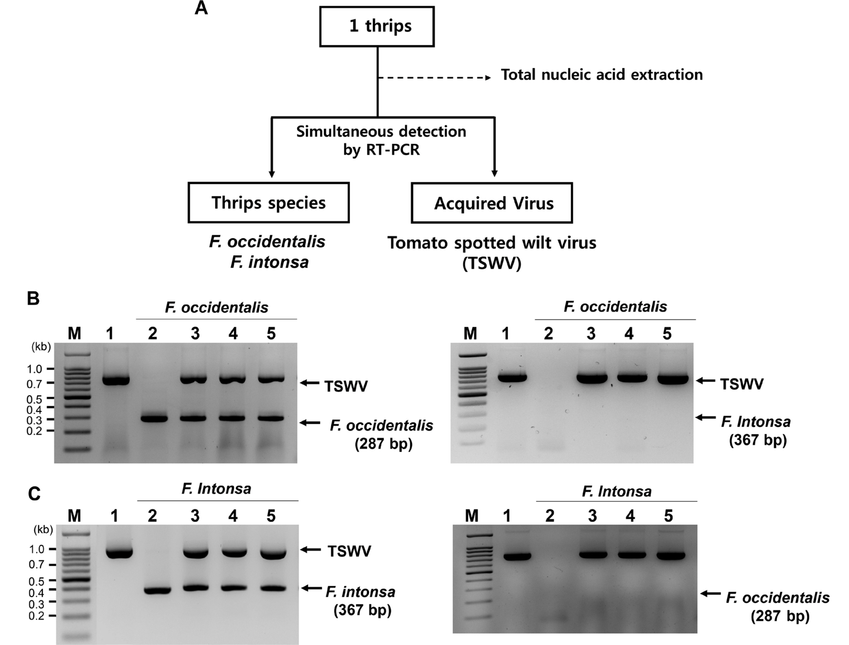
ņ┤Øņ▒äļ▓īļĀłņØś ņóģ ļÅÖņĀĢ ļ░Å ļ│┤ļÅģĒĢśĻ│Ā ņ׳ļŖö TSWVņØś ļŗ©ļÅģ Ēś╣ņØĆ ļÅÖņŗ£ņ¦äļŗ©ņØś Ļ░£ļץņĀü ļ░®ļ▓ĢņØĆ Fig. 2AņŚÉ ņĀ£ņŗ£ļÉśņŚłļŗż. ņ┤Øņ▒äļ▓īļĀłņØś ņóģ ļÅÖņĀĢ ļ░Å ļ│┤ļÅģĒĢśĻ│Ā ņ׳ļŖö TSWVņØś ļŗ©ļÅģ Ēś╣ņØĆ ļÅÖņŗ£ņ¦äļŗ©ņØĆ ĒĢĄņé░ ņČöņČ£ņØä ņØ┤ņÜ®ĒĢśņŚ¼ ĻĮāļģĖļ×æņ┤Øņ▒äļ▓īļĀł ļ░Å ļīĆļ¦īņ┤Øņ▒äļ▓īļĀł ITS2 ļČĆņ£äņŚÉ ĒŖ╣ņØ┤ņĀüņØĖ ĒöäļØ╝ņØ┤ļ©Ė(3ŌĆ▓ ļ¦Éļŗ©ļČĆņŚÉ ņŚŁļ░®Ē¢ź ĒöäļØ╝ņØ┤ļ©ĖļĪ£ņä£ Ļ│ĄĒåĄņĀüņ£╝ļĪ£ ThripsITS2R3ņØ┤ ņé¼ņÜ®ļÉśņŚłņ£╝ļ®░ ĻĮāļģĖļ×æņ┤Øņ▒äļ▓īļĀłļŖö OCC-ITS2F6, ļīĆļ¦īņ┤Øņ▒äļ▓īļĀłļŖö INT-ITSF1ņØä ņĀĢļ░®Ē¢ź ĒöäļØ╝ņØ┤ļ©ĖļĪ£ ņé¼ņÜ®)ļź╝ ņØ┤ņÜ®ĒĢśņŚ¼ RT-PCRļĪ£ ņłśĒ¢ēĒĢśņśĆļŗż(Nakahara ņÖĆ Minoura, 2015). ĻĄŁĒÖö ļ░Å ņ┤Øņ▒äļ▓īļĀłņŚÉņä£ TSWVļź╝ ņ¦äļŗ©ĒĢśĻĖ░ ņ£äĒĢśņŚ¼ TSWVņØś S RNAņŚÉ ņĢöĒśĖĒÖöļÉśņ¢┤ ņ׳ļŖö ņÖĖĒö╝ļŗ©ļ░▒ņ¦ł ņ£ĀņĀäņ×É(N ņ£ĀņĀäņ×É)ļź╝ ĒŖ╣ņØ┤ņĀüņ£╝ļĪ£ ņ”ØĒÅŁĒĢśļÅäļĪØ Ļ│ĀņĢłļÉ£ ĒöäļØ╝ņØ┤ļ©Ė(TSWV-NCP-For ļ░Å TSWV-NCP-Rev)ļź╝ ņØ┤ņÜ®ĒĢśņśĆļŗż(Table 1) (Yoon ļō▒, 2014).
ņČöņČ£ļÉ£ ĒĢĄņé░ 10 ngĻ│╝ TSWV ņ¦äļŗ© ĒöäļØ╝ņØ┤ļ©Ė ņäĖĒŖĖ(TSWV-NCP-For ļ░Å TSWV-NCP-Rev) ļ░Å ņ┤Øņ▒äļ▓īļĀł ņ¦äļŗ© ĒöäļØ╝ņØ┤ļ©Ė ņäĖĒŖĖ(OCC-ITS2F6ņÖĆ ThripsITSR3 Ēś╣ņØĆ INT-ITSF1 ļ░Å ThripsITSR3)ļź╝ 10 pmolņö® ļ░śņØæņĢĪņŚÉ ļäŻĻ│Ā ņĀ£ņĪ░ĒÜīņé¼ņØś ņŗżĒŚśļ░®ļ▓ĢņŚÉ ļö░ļØ╝ SuPrimeScript RT-PCR premix (GeNetBio, Nonsan, Korea)ļź╝ ņØ┤ņÜ®ĒĢśņŚ¼ ļ░śņØæņŗ£ņ╝░ļŗż. ĻĄÉņ░©ļ░śņØæņØä ĒÖĢņØĖĒĢśĻĖ░ ņ£äĒĢ┤ ĻĮāļģĖļ×æņ┤Øņ▒äļ▓īļĀłņŚÉņä£ ņČöņČ£ĒĢ£ ĒĢĄņé░ņŚÉ ļīĆļ¦īņ┤Øņ▒äļ▓īļĀł ņ¦äļŗ©ņØä ņ£äĒĢ£ ĒöäļØ╝ņØ┤ļ©Ė(INT-ITSF1 ļ░Å ThripsITSR3)ļź╝ ļäŻņŚłĻ│Ā, ļīĆļ¦īņ┤Øņ▒äļ▓īļĀłņŚÉņä£ ņČöņČ£ĒĢ£ ĒĢĄņé░ņŚÉļŖö ĻĮāļģĖļ×æņ┤Øņ▒äļ▓īļĀł ņ¦äļŗ©ņØä ņ£äĒĢ£ ĒöäļØ╝ņØ┤ļ©Ė(OCC-ITS2F6ņÖĆ ThripsITSR3)ļź╝ ļäŻņ¢┤ RT-PCRņØä ņłśĒ¢ēĒĢśņśĆļŗż. RT-PCR ņĪ░Ļ▒┤ņØĆ 50┬░CņŚÉņä£ 50ļČä(1ĒÜī) ļÅÖņĢł ņŚŁņĀäņé¼ ļ░śņØæņŚÉ ņØ┤ņ¢┤ 95┬░CņŚÉņä£ 2ļČä(1ĒÜī), 95┬░C 15ņ┤ł, 55┬░C 30ņ┤ł, 72┬░C, 50ņ┤ł(35ĒÜī ļ░śļ│Ą), 72┬░CņŚÉņä£ 5ļČä(1ĒÜī)ņØ┤ņŚłļŗż. ņĀäĻĖ░ņśüļÅÖņØĆ 0.5├ŚTBE ņÖäņČ®ņĢĪņÖĆ 1.0% ņĢäĻ░ĆļĪ£ņŖż ņĀżņŚÉņä£ ņŗżņŗ£ĒĢśņśĆĻ│Ā, DNA ņé¼ņØ┤ņ”ł ļ¦łņ╗żļĪ£ HiQ 100 bp plus DNA ladder (BioD, Gwangmyeong, Korea)ļź╝ ņØ┤ņÜ®ĒĢśņśĆļŗż. PCR ņ”ØĒÅŁ ņé░ļ¼╝ņØĆ 110ļ│╝ĒŖĖņŚÉņä£ 50ļČä ļÅÖņĢł ņĀäĻĖ░ņśüļÅÖ Ēøä UV transilluminatorļź╝ ņØ┤ņÜ®ĒĢśņŚ¼ Ļ▓░Ļ│╝ļź╝ ĒÖĢņØĖĒĢśņśĆļŗż.
Ļ▓░Ļ│╝ ļ░Å Ļ│Āņ░░
ĻĄŁĒÖöņŚÉņä£ TSWV Ēö╝ĒĢ┤ ņĪ░ņé¼
2019ļģä ĻĄŁĒÖö ņŻ╝ņÜö ņ×¼ļ░░ņ¦ĆņŚŁ ņżæ ĒĢśļéśņØĖ Ļ▓Įļé© ņ░ĮņøÉņŗ£ ņåīņ×¼ ņŗ£ņäżĒĢśņÜ░ņŖżņŚÉņä£ ņ×¼ļ░░ļÉśļŖö ņŖżĒāĀļŗżļō£ ĻĄŁĒÖö(ļ░▒ļ¦ł ĒÆłņóģ)ļź╝ ņĪ░ņé¼ĒĢ£ Ļ▓░Ļ│╝, 2ņøöŌĆÆ3ņøö ĻĄŁĒÖö ņĀĢņŗØ ņØ┤Ēøä 4ņøöĻ▓Į ņ┤Øņ▒äļ▓īļĀłĻ░Ć ĒĢśņÜ░ņŖż ņŗ£ņäż ļé┤ļĪ£ ņ£Āņ×ģļÉśņ¢┤ ņ┤Øņ▒äļ▓īļĀłĻ░Ć TSWV Ļ░ÉņŚ╝ ĻĄŁĒÖöņŚÉņä£ TSWVļź╝ ņ▓┤ļé┤ ļ│┤ļÅģ Ēøä ņ¦ĆņåŹņĀüņ£╝ļĪ£ TSWV Ļ░ÉņŚ╝ņØä ĒÖĢņé░ņŗ£ĒéżļŖö Ļ▓āņ£╝ļĪ£ ĒÖĢņØĖļÉśņŚłļŗż. 5ņøöļČĆĒä░ ņ£ĪņĢł ĒÖĢņØĖņØ┤ Ļ░ĆļŖźĒĢ£ TSWV ļ│æņ¦ĢņØ┤ ļéśĒāĆļéśĻĖ░ ņŗ£ņ×æĒ¢łļŗż. ļ│æņ¦Ģ ņ┤łĻĖ░ņŚÉļŖö Ļ░ÉņŚ╝ ĻĄŁĒÖö ņ×ÄņØ┤ ĒŗĆņ¢┤ņ¦ĆĻ▒░ļéś ņżäĻĖ░Ļ░Ć Ē£śņ¢┤ņ¦ĆļŖö ļ│æņ¦ĢņØ┤ ļéśĒāĆļé¼ņ£╝ļ®░, ļ│æņØ┤ ņ¦äņĀäļÉśļ®┤ņä£ ņāüņŚĮņŚÉ Ļ┤┤ņé¼ļ░śņĀÉĻ│╝ ņżäĻĖ░ Ļ┤┤ņé¼Ļ░Ć ļéśĒāĆļéś Ļ┤┤ņé¼ļÉ£ ļ░®Ē¢źņ£╝ļĪ£ ņŗØļ¼╝ņ▓┤Ļ░Ć ĻĖ░ņÜĖņ¦ĆļŖö ļ│æņ¦ĢņØ┤ Ļ┤Ćņ░░ļÉśņŚłļŗż. Ļ░ÉņŚ╝ ņØ┤Ēøä 2Ļ░£ņøö ņØ┤ļé┤ņŚÉ ņĀĢļŗ©ļČĆ ņżäĻĖ░Ļ░Ć Ļ┤┤ņé¼ļÉśļ®┤ņä£ ņāüņŚĮņØ┤ Ļ│Āņé¼ļÉśļŖö ņ┤łĻĖ░ ļ│æņ¦ĢņØ┤ ļéśĒāĆļé¼Ļ│Ā ņØ┤Ēøä ņŗØļ¼╝ņ▓┤Ļ░Ć ņĀäņ▓┤Ļ░Ć Ļ│Āņé¼Ē¢łļŗż(Fig. 1A). TSWVņŚÉ ņØśĒĢ£ Ļ░ÉņŚ╝ņŚ¼ļČĆļź╝ ĒÖĢņØĖĒĢśĻĖ░ ņ£äĒĢ┤ Ļ░ÉņŚ╝ ņ×ÄņØä ļ¦łņćäĒĢ£ ņĪ░ņ”ÖņĢĪņŚÉ Im-munoStrip (Agdia)ļź╝ ļŗ┤Ļ░Ć ĒÖĢņØĖĒĢ£ Ļ▓░Ļ│╝, TSWV ĒĢŁņ▓┤-ĻĖł ņ×ģņ×ÉĻ░Ć TSWVņÖĆ ļ░śņØæĒĢ£ ļČēņØĆ ņäĀņØ┤ 2Ļ░£Ļ░Ć ļéśĒāĆļéś TSWV Ļ░ÉņŚ╝ņØ┤ ĒÖĢņ¦äļÉśņŚłļŗż(Fig. 1B). ņŚŁĒĢÖ ņĪ░ņé¼ Ļ▓░Ļ│╝, ĻĄŁĒÖö ņŗ£ņäżĒĢśņÜ░ņŖżņØś ņĖĪņ░Į ņŻ╝ļ│ĆņŚÉ ņŗ¼ņØĆ ĻĄŁĒÖöņŚÉņä£ Ļ░Ćņן ļ©╝ņĀĆ TSWV ņ”ØņāüņØ┤ ļéśĒāĆļéśļŖö Ļ▓āņ£╝ļĪ£ ĒÖĢņØĖļÉśņŚłņ£╝ļ®░ ņ┤Øņ▒äļ▓īļĀłņØś ņ”ØĻ░ĆņÖĆ ĒĢ©Ļ╗ś ņŗ£ņäżĒĢśņÜ░ņŖż Ļ░Ćņן ņĢłņ¬ĮņØś ĻĄŁĒÖöĻ╣īņ¦Ć ĒÖĢņé░ļÉśļŖö Ļ▓āņ£╝ļĪ£ ĒÖĢņØĖļÉśņŚłļŗż(data not shown).
ņ┤Øņ▒äļ▓īļĀł ņóģ ļÅÖņĀĢ ļ░Å ņ┤Øņ▒äļ▓īļĀł ļ│┤ļÅģ TSWV ļÅÖņŗ£ņ¦äļŗ©ļ▓Ģ Ļ░£ļ░£
TSWVņØś ĒŖ╣ņØ┤ņĀüņØĖ ĒöäļØ╝ņØ┤ļ©Ėļź╝ ņØ┤ņÜ®ĒĢśņŚ¼ Ļ│ĀņČöņŚÉņä£ ņ”ØņŗØļÉ£ TSWVļź╝ ņ¦äļŗ©ĒĢ£ Ļ▓░Ļ│╝ 777 bpĻ░Ć ĒÖĢņØĖļÉśņŚłļŗż(Supplementary Fig. 1A). ļśÉĒĢ£ ĻĄŁĒÖö ņŗ£ņäżĒĢśņÜ░ņŖż ņŻ╝ļ│ĆņŚÉņä£ ņ▒äņ¦æĒĢ£ ĻĮāļģĖļ×æņ┤Øņ▒äļ▓īļĀł ļ░Å ļīĆļ¦īņ┤Øņ▒äļ▓īļĀłļź╝ ĒśĢĒā£ņĀüņ£╝ļĪ£ ĻĄ¼ļČäĒĢ£ Ēøä Ļ░ü 10ļ¦łļ”¼ņö®ņØä ļ¬©ņĢäņä£ ĒĢĄņé░ņØä ņČöņČ£ĒĢ£ Ēøä ĒŖ╣ņØ┤ ĒöäļØ╝ņØ┤ļ©Ėļź╝ ņØ┤ņÜ®ĒĢśņŚ¼ RT-PCRņØä ņłśĒ¢ēĒĢ£ Ļ▓░Ļ│╝, ĻĮāļģĖļ×æņ┤Øņ▒äļ▓īļĀłļŖö 287 bp, ļīĆļ¦īņ┤Øņ▒äļ▓īļĀłļŖö 367 bpņØś DNAĻ░Ć ĻĄÉņ░©ļ░śņØæ ņŚåņØ┤ ņ”ØĒÅŁļÉśņŚłļŗż(data not shown). ņ┤Øņ▒äļ▓īļĀł 1 ļ¦łļ”¼(0.05 mg)ņŚÉņä£ ņ┤Øņ▒äļ▓īļĀłņŚÉņä£ ņóģ ĻĄ¼ļČä ļ░Å ņ▓┤ļé┤ņŚÉ ļ│┤ļÅģĒĢśĻ│Ā ņ׳ļŖö TSWVļź╝ ļÅÖņŗ£ņ¦äļŗ©ĒĢśĻĖ░ ņ£äĒĢ┤ ĒĢĄņé░ņØä ņČöņČ£ĒĢśņśĆņ£╝ļ®░, ņĀĢļ¤ēĒĢ£ Ļ▓░Ļ│╝ 100ŌĆÆ200 ngņØś ĒĢĄņé░ņØ┤ ņČöņČ£ļÉśņŚłļŗż. 10ŌĆÆ20 ng ĒĢĄņé░ņØä ņØ┤ņÜ®ĒĢśņŚ¼ ņĢ×ņä£ ĻĖ░ņłĀĒĢ£ ņ┤Øņ▒äļ▓īļĀłņØś ņóģ ĒŖ╣ņØ┤ņĀü ĒöäļØ╝ņØ┤ļ©Ėļź╝ ņØ┤ņÜ®ĒĢśņŚ¼ RT-PCRņØä ņłśĒ¢ēĒĢ£ Ļ▓░Ļ│╝ ņ”ØĒÅŁņé░ļ¼╝ņØ┤ ļ╣äĒŖ╣ņØ┤ņĀü ļ░śņØæ ņŚåņØ┤ ņ”ØĒÅŁļÉśņŚłļŗż(Supplementary Fig. 1B, C). ĻĄŁĒÖö ņŗ£ņäżņ×¼ļ░░ņ¦ĆņŚÉņä£ Ēżņ¦æĒĢ£ ĻĮāļģĖļ×æņ┤Øņ▒äļ▓īļĀł ņä▒ņČ®ļ┐Éļ¦ī ņĢäļŗłļØ╝ ĻĮāļģĖļ×æņ┤Øņ▒äļ▓īļĀł 1ļĀ╣ ņĢĮņČ®ņŗ£ĻĖ░ņŚÉ TSWVļź╝ ĒØĪņ”Öņŗ£Ēé© Ēøä ņé¼ņ£ĪĒĢ£ 2ļĀ╣ ņĢĮņČ®Ļ│╝ ļ▓łļŹ░ĻĖ░ 1ļ¦łļ”¼ņŚÉņä£ TSWVĻ░Ć ņ¦äļŗ©ļÉśņŚłļŗż(Supplementary Fig. 2).
ņØĖņ£äņĀüņ£╝ļĪ£ TSWVļź╝ ļ│┤ļÅģņŗ£Ēé© F. occidentalis ļ░Å ļīĆļ¦īņ┤Øņ▒äļ▓īļĀłņØś ņä▒ņČ® 1ļ¦łļ”¼ņŚÉņä£ TSWVņÖĆ ņ┤Øņ▒äļ▓īļĀł ņóģ ļÅÖņĀĢņØä ļÅÖņŗ£ ņ¦äļŗ©ĒĢ£ Ļ▓░Ļ│╝, ĻĮāļģĖļ×æņ┤Øņ▒äļ▓īļĀł ļ░Å ļīĆļ¦īņ┤Øņ▒äļ▓īļĀł ĻĄÉņ░© ļ░śņØæņØ┤ ņŚåņØ┤ TSWVņÖĆ ņ┤Øņ▒äļ▓īļĀł ņóģ ĒŖ╣ņØ┤ņĀüņØĖ ņ”ØĒÅŁ ņé░ļ¼╝ņØ┤ ĒĢ®ņä▒ņØ┤ ļÉśņ¢┤ ļÅÖņŗ£ņ¦äļŗ©ņØ┤ Ļ░ĆļŖźĒĢ£ Ļ▓āņ£╝ļĪ£ ĒīÉņĀĢļÉśņŚłļŗż(Fig. 2B, C). ĻĖ░ņĪ┤ņØś ņ┤Øņ▒äļ▓īļĀłņØś ņóģņØä ĻĄ¼ļČäĒĢśĻĖ░ ņ£äĒĢ£ ļČäņ×Éņ¦äļŗ©ņØĆ ļ»ĖĒåĀņĮśļō£ļ”¼ņĢä DNA cytochrome oxidase subunit I ļČĆļČäņØä PCRļĪ£ ņ”ØĒÅŁĒĢ£ ļŗżņØī ņĀ£ĒĢ£ĒÜ©ņåī RsaIņ£╝ļĪ£ ņĀłļŗ©ĒĢśņŚ¼ ļéśĒāĆļé£ DNA ļŗ©ĒÄĖņØś Ēī©Ēä┤ņ£╝ļĪ£ ĻĄ¼ļČäĒĢśļŖö ļ░®ļ▓ĢņØ┤ļéś(Brunner ļō▒, 2002) Ēś╣ņØĆ ribosomal ITS2 ņ£ĀņĀäņ×Éļź╝ PCRļĪ£ ņ”ØĒÅŁ Ēøä ņ┤Øņ▒äļ▓īļĀłņØś Ļ░ü ņóģļ¦łļŗż ņä£ļĪ£ ļŗżļźĖ ņĀ£ĒĢ£ĒÜ©ņåīļź╝ ņ▓śļ”¼ĒĢśņŚ¼ restriction fragment length polymorphism Ļ▓░Ļ│╝ļĪ£ ĒīÉņĀĢĒĢśļŖö ļ░®ļ▓ĢņØä ņé¼ņÜ®ĒĢśņśĆļŗż(Brunner ļō▒, 2002; TodaņÖĆ Komazaki, 2002). ņĄ£ĻĘ╝ņŚÉļŖö ļō▒ņś©ņ”ØĒÅŁļ▓Ģ(loop-mediated isothermal amplification) ļ░Å recombinase polymerase amplification ņ¦äļŗ©ļ▓ĢņØ┤ Ļ░£ļ░£ļÉśņŚłļŗż(Fekrat ļō▒, 2015; Fukuda ļō▒, 2005; Priti ļō▒, 2020; Przybylska ļō▒, 2015). ĻĘĖļ¤¼ļéś ņ▓śļ”¼ĒĢśļŖö ļŹ░ ņŗ£Ļ░äņØ┤ ļ¦ÄņØ┤ ņåīņÜöļÉśļ®░ PCR ļ░śņØæņØ┤ ļüØļé£ ĒøäņŚÉļÅä ņĀ£ĒĢ£ĒÜ©ņåīļź╝ ņČöĻ░ĆļĪ£ ņ▓śļ”¼ĒĢśĻ▒░ļéś, ņ×¼Ēśäņä▒ņØ┤ ļ¢©ņ¢┤ņ¦ĆĻ▒░ļéś, ĒöäļØ╝ņØ┤ļ©Ė ņĀ£ņ×æņŚÉ ļ│Ąņ×ĪĒĢ£ ņĀłņ░©Ļ░Ć ĒĢäņÜöĒĢśļŗżļŖö ļŗ©ņĀÉņØ┤ ņ׳ņŚłļŗż. ņØ┤ļ▓ł ņŚ░ĻĄ¼ņŚÉņä£ Ļ░£ļ░£ļÉ£ ņ┤Øņ▒äļ▓īļĀł ļ░Å TSWV ļÅÖņŗ£ ļŗżņżæ PCR ņ¦äļŗ©ļ▓ĢņØĆ ĻĄŁĒÖö ņŗ£ņäżĒĢśņÜ░ņŖżņŚÉņä£ ņÖĖļČĆ ņ£Āņ×ģ ņ┤Øņ▒äļ▓īļĀłņØś ļ░öņØ┤ļ¤¼ņŖż ļ│┤ļÅģ ņŚ¼ļČĆļź╝ ņēĮĻ│Ā ļ╣Āļź┤Ļ▓ī ņ¦äļŗ©ĒĢĀ ņłś ņ׳ĻĖ░ ļĢīļ¼ĖņŚÉ ļ░öņØ┤ļ¤¼ņŖżļ│æ ņ£Āņ×ģ ļ░Å ĒÖĢņé░ņØä ņśłņĖĪĒĢśļŖö ļŹ░ ĒÖ£ņÜ®ĒĢĀ ņłś ņ׳ņØä Ļ▓āņ£╝ļĪ£ ĒīÉļŗ©ļÉ£ļŗż(Jung ļō▒ 2019; Kang ļō▒, 2012).
ņŗ£ņäżņ×¼ļ░░ ĻĄŁĒÖöņŚÉņä£ ņłśņ¦æļÉ£ ņ┤Øņ▒äļ▓īļĀłņØś TSWV ļ│┤ļÅģļźĀ ņĪ░ņé¼
ņ┤Øņ▒äļ▓īļĀł 1ļ¦łļ”¼ņŚÉņä£ TSWV ļ│┤ļÅģ ņŚ¼ļČĆ ļ░Å ņóģ ļÅÖņĀĢņØ┤ Ļ░ĆļŖźĒĢ£ Ļ▓░Ļ│╝ņØś ĒÜ©ņÜ®ņä▒ņØä ņĪ░ņé¼ĒĢśĻĖ░ ņ£äĒĢśņŚ¼, ĻĄŁĒÖö ņ×¼ļ░░ ĒĢśņÜ░ņŖżņŚÉņä£ ņ┤Øņ▒äļ▓īļĀłņÖĆ TSWVļź╝ ļÅÖņŗ£ ņ¦äļŗ©ņØä ņŗżņŗ£ĒĢśņśĆļŗż. ņØ┤ļ¤░ ļ¬®ņĀüņ£╝ļĪ£ ņČ®ļé© Ēā£ņĢłĻĄ░ ņåīņ×¼ ņŖżĒöäļĀłņØ┤ ĻĄŁĒÖö(ņåīĻĄŁ) ņ×¼ļ░░ ĒĢśņÜ░ņŖż ļ░Å Ļ▓Įļé© ņ░ĮņøÉņŗ£ ņåīņ×¼ ņŖżĒāĀļŗżļō£ ĻĄŁĒÖö(ļīĆĻĄŁ) ņ×¼ļ░░ ĒĢśņÜ░ņŖżņŚÉņä£ ĒāĆļØĮļ▓ĢņØä ņØ┤ņÜ®ĒĢśņŚ¼ Ļ░üĻ░ü 43, 102ļ¦łļ”¼ļź╝ ļ¼┤ņ×æņ£äļĪ£ ņ▒äņ¦æĒĢ£ ļŗżņØī ļÅÖņŗ£ņ¦äļŗ©ļ▓Ģņ£╝ļĪ£ ņóģ ļÅÖņĀĢ ļ░Å TSWV ļ│┤ļÅģ ņŚ¼ļČĆļź╝ ņĪ░ņé¼ĒĢśņśĆļŗż.
Ēā£ņĢłĻĄ░ņŚÉņä£ ņ▒äņ¦æĒĢ£ ņ┤Øņ▒äļ▓īļĀłņŚÉņä£ ĻĮāļģĖļ×æņ┤Øņ▒äļ▓īļĀłļŖö 36ļ¦łļ”¼ļĪ£ 83.7%ļź╝ ņ░©ņ¦ĆĒ¢łņ£╝ļ®░, ļÅÖņĀĢļÉ£ ĻĮāļģĖļ×æņ┤Øņ▒äļ▓īļĀłņŚÉņä£ 36ļ¦łļ”¼ ņżæ 27ļ¦łļ”¼ņŚÉņä£ TSWVĻ░Ć ņ¦äļŗ©ļÉśņ¢┤, ĻĮāļģĖļ×æņ┤Øņ▒äļ▓īļĀłņØś TSWV ļ│┤ļÅģļźĀņØĆ 72.9%ļĪ£ ņĪ░ņé¼ļÉśņŚłļŗż(Table 2). ņóģ ļÅÖņĀĢ Ļ▓░Ļ│╝ ļīĆļ¦īņ┤Øņ▒äļ▓īļĀłļŖö 1ļ¦łļ”¼ļĪ£ ĒīÉņĀĢļÉśņŚłņ£╝ļ®░, TSWVļŖö ļ│┤ļÅģĒĢśĻ│Ā ņ׳ņ¦Ć ņĢŖņĢśņ£╝ļ®░, ļ»ĖļÅÖņĀĢ ņ┤Øņ▒äļ▓īļĀłļŖö 43ļ¦łļ”¼ ņżæ 6ļ¦łļ”¼ņØ┤ņŚłņ£╝ļ®░, TSWV ļ│┤ļÅģļźĀņØĆ 100% (6ļ¦łļ”¼ ņżæ 6ļ¦łļ”¼)ļĪ£ ņĪ░ņé¼ļÉśņŚłļŗż(Table 2).
Ļ▓Įļé© ņ░ĮņøÉņŗ£ņŚÉņä£ ņ▒äņ¦æĒĢ£ ņ┤Øņ▒äļ▓īļĀł ņóģ ļÅÖņĀĢ ļ░Å TSWV ļ│┤ļÅģļźĀņØä ņĪ░ņé¼ĒĢ£ Ļ▓░Ļ│╝, 102 ļ¦łļ”¼ ņżæ 94ļ¦łļ”¼(92.2%)Ļ░Ć ĻĮāļģĖļ×æņ┤Øņ▒äļ▓īļĀłļĪ£ ĒÖĢņØĖļÉśņŚłņ£╝ļ®░, 94ļ¦łļ”¼ ĻĮāļģĖļ×æņ┤Øņ▒äļ▓īļĀł ņżæ 79 ļ¦łļ”¼ņŚÉņä£ TSWVĻ░Ć Ļ▓ĆņČ£ļÉśņ¢┤ TSWV ļ│┤ļÅģļźĀņØĆ 84.0%ļĪ£ ņĪ░ņé¼ļÉśņŚłļŗż(Table 2). ļīĆļ¦īņ┤Øņ▒äļ▓īļĀłļź╝ ļÅÖņĀĢĒĢ£ Ļ▓░Ļ│╝ 102ļ¦łļ”¼ ņżæ 4ļ¦łļ”¼(3.9%)ņØ┤ņŚłņ£╝ļ®░ TSWV ļ│┤ļÅģļźĀņØĆ 50%ļĪ£ ņĪ░ņé¼ļÉśņŚłļŗż(Table 2). ĻĮāļģĖļ×æņ┤Øņ▒äļ▓īļĀł ļ░Å ļīĆļ¦īņ┤Øņ▒äļ▓īļĀł ņØ┤ņÖĖņŚÉ ļŗżļźĖ ņóģņØś ņ┤Øņ▒äļ▓īļĀłļŖö 102ļ¦łļ”¼ ņżæ 4ļ¦łļ”¼(3.0%) ņØ┤ņŚłņ£╝ļ®░, TSWV ļ│┤ļÅģļźĀņØĆ 75%ļĪ£ ņĪ░ņé¼ļÉśņŚłļŗż. ņĪ░ņé¼ļÉ£ ņé¼ņŗżņØä ņóģĒĢ®ĒĢ┤ļ│╝ ļĢī, ĻĄŁĒÖö ņŗ£ņäż ĒĢśņÜ░ņŖżņŚÉņä£ ņÜ░ņĀÉņóģņØĆ ĻĮāļģĖļ×æņ┤Øņ▒äļ▓īļĀłņØ┤ļ®░ 75.0ŌĆÆ84.0% TSWV ļ│┤ļÅģļźĀņØä Ļ░Ćņ¦ĆĻ│Ā ņ׳ņ¢┤ TSWV Ļ░ÉņŚ╝ ļ░Å ņĀäĒīīņŚÉ Ļ░Ćņן Ēü░ ņÜöņØĖņ×äņØä ĒīÉļŗ©ļÉ£ļŗż. ĒØźļ»ĖļĪŁĻ▓īļÅä ļ╣äļĪØ ņ┤Øņ▒äļ▓īļĀł ņóģ ļÅÖņĀĢņØĆ ĒÖĢņØĖĒĢĀ ņłś ņŚåņŚłņ£╝ļéś ļ»ĖļÅÖņĀĢ ņ┤Øņ▒äļ▓īļĀłņŚÉņä£ TSWV ļ│┤ļÅģļźĀņØĆ ņāüļŗ╣Ē׳ ļåÆņØĆ ņłśņżĆņ£╝ļĪ£ ņĪ░ņé¼ļÉśņŚłļŗż. TSWVļź╝ ļ¦żĻ░£ĒĢĀ ņłś ņ׳ļŗżĻ│Ā ņĢīļĀżņ¦ä ņśżņØ┤ņ┤Øņ▒äļ▓īļĀł ņŚ¼ļČĆļź╝ ļ»ĖļÅÖņĀĢļÉ£ ņ┤Øņ▒äļ▓īļĀłļź╝ Ēśäļ»ĖĻ▓ĮņŚÉņä£ ĒśĢĒā£ĒĢÖņĀü ļČäļźśļź╝ ĒĢśņśĆņ£╝ļéś ņśżņØ┤ņ┤Øņ▒ä ļ▓īļĀłļŖö ĒÖĢņØĖļÉśņ¦Ć ņĢŖņĢśļŗż(data not shown). ĻĘĖ ņÖĖ ĻĄŁĒÖöņŚÉņä£ ņä£ņŗØĒĢśļŖö ņ┤Øņ▒äļ▓īļĀłļŖö Ēīīņ┤Øņ▒äļ▓īļĀł, ĒĢśņÖĆņØ┤ņ┤Øņ▒äļ▓īļĀł, ļ»Ėļéśļ”¼ņ┤Øņ▒äļ▓īļĀł ļō▒ 10ņŚ¼ņóģņØ┤ ĒÖĢņØĖļÉśĻ│Ā ņ׳ņ£╝ļéś TSWV ļ│┤ļÅģ ņŚ¼ļČĆ ļō▒ņŚÉ ļīĆĒĢśņŚ¼ ļ®┤ļ░ĆĒĢ£ ņĪ░ņé¼Ļ░Ć ĒĢäņÜöĒĢśļŗżĻ│Ā ĒīÉļŗ©ļÉ£ļŗż(van de Wetering, 1999).
ņÜö ņĢĮ
ņØ┤ļ▓ł ņŚ░ĻĄ¼ļŖö ĻĄŁĒÖöņŚÉņä£ ļ¼ĖņĀ£ļÉśļŖö ņ┤Øņ▒äļ▓īļĀłņØś ņóģ ļÅÖņĀĢ ļ░Å ļ│┤ļÅģ ļ░öņØ┤ļ¤¼ņŖżņØĖ ĒåĀļ¦łĒåĀļ░śņĀÉņ£äņĪ░ļ░öņØ┤ļ¤¼ņŖż(Tomato spotted wilt virus, TSWV)ļź╝ ļÅÖņŗ£ņŚÉ ĒÖĢņØĖĒĢĀ ņłś ņ׳ļŖö ņ¦äļŗ©ļ░®ļ▓ĢņØä Ļ░£ļ░£ĒĢśņśĆļŗż. ņØ┤ļŖö ņ┤Øņ▒äļ▓īļĀł 1ļ¦łļ”¼ņŚÉņä£ ņČöņČ£ĒĢ£ ĒĢĄņé░ņŚÉ ĻĮāļģĖļ×æņ┤Øņ▒äļ▓īļĀł ļ░Å ļīĆļ¦īņ┤Øņ▒äļ▓īļĀłņØś ITS2 ļČĆļČäņŚÉ ĒŖ╣ņØ┤ņĀüņØĖ ĒöäļØ╝ņØ┤ļ©ĖņÖĆ TSWV ņÖĖĒö╝ļŗ©ļ░▒ņ¦ł(N) ņ£ĀņĀäņ×É ĒŖ╣ņØ┤ņĀüņØĖ ĒöäļØ╝ņØ┤ļ©Ėļź╝ ļÅÖņŗ£ņŚÉ ļäŻņ¢┤ reverse tran scriptionŌĆÆpolymerase chain reactionņØä ņłśĒ¢ēĒĢśņŚ¼ DNAļź╝ ņ”ØĒÅŁņŗ£ĒéżļŖö ļ░®ļ▓Ģņ£╝ļĪ£ ņĀäĻĖ░ņśüļÅÖĒĢśņŚ¼ Ļ░üĻ░ü 287, 367, 777 bpņØś DNA ļŗ©ĒÄĖņØś Ēü¼ĻĖ░ļź╝ ļ╣äĻĄÉĒĢ©ņ£╝ļĪ£ņŹ© ņ┤Øņ▒äļ▓īļĀłņØś ņóģ ļÅÖņĀĢ ļ░Å ņ┤Øņ▒äļ▓īļĀłņØś TSWV ļ│┤ļÅģ ņŚ¼ļČĆļź╝ ļÅÖņŗ£ņŚÉ ĒÖĢņØĖĒĢĀ ņłś ņ׳ļŗż. ņČ®ņ▓Łļé©ļÅä Ēā£ņĢł ļ░Å Ļ▓Įņāüļé©ļÅä ņ░ĮņøÉņØś ĻĄŁĒÖö ņŗ£ņäżĒĢśņÜ░ņŖżņŚÉņä£ ņ┤Øņ▒äļ▓īļĀłļź╝ Ēżņ¦æĒĢśņŚ¼ ņ┤Øņ▒äļ▓īļĀł ņÜ░ņĀÉņóģĻ│╝ ņ┤Øņ▒äļ▓īļĀłņØś TSWV ļ│┤ļÅģņ£©ņØä ņĪ░ņé¼ĒĢ£ Ļ▓░Ļ│╝, Ēā£ņĢłņØś ĻĄŁĒÖö ņŗ£ņäżĒĢśņÜ░ņŖżņŚÉņä£ļŖö ĻĮāļģĖļ×æņ┤Øņ▒äļ▓īļĀłĻ░Ć 83.7%ļĪ£ ņÜ░ņĀÉĒĢśĻ│Ā ņ׳ņ£╝ļ®░ ņ▒äņ¦æļÉ£ ņ┤Øņ▒äļ▓īļĀł ņżæ 72.9%Ļ░Ć TSWVļź╝ ļ│┤ļÅģĒĢśĻ│Ā ņ׳ņŚłņ£╝ļ®░, ņ░ĮņøÉņŚÉņä£ļŖö ĻĮāļģĖļ×æņ┤Øņ▒äļ▓īļĀłĻ░Ć 92.2%ļź╝ ņ░©ņ¦ĆĒĢśĻ│Ā ņ׳ņ£╝ļ®░ 84.0%ņØś ņ┤Øņ▒äļ▓īļĀłņŚÉņä£ TSWVĻ░Ć ņ¦äļŗ©ļÉśņŚłļŗż. ņØ┤ļ¤¼ĒĢ£ Ļ▓░Ļ│╝ļŖö Frankliniella occidentalisĻ░Ć ņÜ░ņĀÉņóģņØ┤ļ®░ ņś©ņŗżņØś ĻĄŁĒÖö ņŗØļ¼╝ņŚÉņä£ TSWVņØś ņĀäļ░śņŚÉ ņżæņÜöĒĢ£ ņŚŁĒĢĀņØä ĒĢ£ļŗżļŖö Ļ▓āņØä ĒÖĢņØĖĒĢ┤ņżĆļŗż. ņØ┤ļ▓ł ņŚ░ĻĄ¼ļŖö ĻĄŁĒÖö ņŗ£ņäżĒĢśņÜ░ņŖżņŚÉņä£ ņ┤Øņ▒äļ▓īļĀłļź╝ ĒåĄĒĢ£ TSWVņØś ņŗ£ņäżĒĢśņÜ░ņŖżļé┤ ņ£Āņ×ģņŗ£ĻĖ░ ļ░Å ĒÖĢņé░ Ļ▓ĮļĪ£ ļō▒ ļ░öņØ┤ļ¤¼ņŖżņØś ņŚŁĒĢÖņŚ░ĻĄ¼ļź╝ ņ£äĒĢ£ Ļ░äĒÄĖņ¦äļŗ©ļ▓Ģņ£╝ļĪ£ ĒÖ£ņÜ® Ļ░ĆļŖźĒĢ©ņØä ņśłņŗ£ĒĢ┤ņżĆļŗż.
Electronic Supplementary Material
Supplementary materials are available at Research in Plant Disease website (http://www.online-rpd.org/).
 PDF Links
 PubReader
 ePub Link
 Full text via DOI
 Download Citation
 Supplement
 Print
Print


