

ņä£ļĪĀ
ĒĢ┤ļ░öļØ╝ĻĖ░(Helianthus annuus L.)ļŖö ĻĄŁĒÖöĻ│╝(Asteraceae)ņØś 1ļģäņāØ ņ┤łļ│ĖņŗØļ¼╝ļĪ£ ļČüņĢäļ®öļ”¼ņ╣┤ ņżæņä£ļČĆ┬Ę ņ¦ĆņŚŁņØ┤ ņøÉņé░ņ¦ĆņØ┤ļŗż(Heiser, 1951). ļĢģņĮ®, ņĮ®, ņ£Āņ▒ä ļō▒Ļ│╝ ļŹöļČłņ¢┤ 4ļīĆ ņ£Āņ¦Ćņ×æļ¼╝ ņżæņØś ĒĢśļéśļĪ£ ĒĢ┤ļ░öļØ╝ĻĖ░ņö©ņŚÉļŖö ņ¦Ćļ░®ņ£Ā, ĒĢäņłśņĢäļ»ĖļģĖņé░ņØ┤ ĒÆŹļČĆĒĢśņŚ¼ ĒśłņĢĪņł£ĒÖśņØä ļÅäņÖĆņŻ╝ļ®░ ļ╣äĒāĆļ»╝ ĒĢ©ļ¤ēņØ┤ ļ¦ÄņĢä ņśüņ¢æĒĢÖņĀüņ£╝ļĪ£ ņÜ░ņłśĒĢ£ ņŗØĒÆłņ£╝ļĪ£ ĻČīņןļÉ£ļŗż(BaudetĻ│╝ Moss├®, 1977; ┼Ākori─ć ļō▒, 2008; Warner, 2005).
ņäĖĻ│äņĀüņ£╝ļĪ£ ĒĢ┤ļ░öļØ╝ĻĖ░ņŚÉņä£ ļ░£ļ│æļÉśļŖö ņŻ╝ņÜö ļ│æņøÉņ£╝ļĪ£ļŖö ĒÆŗļ¦łļ”äļ│æ(Ralstonia solanacearum), Ļ▓ĆņØĆļ¼┤ļŖ¼ļ│æ(Alternaria spp.), ņ×┐ļ╣øĻ│░ĒīĪņØ┤ļ│æ(Botrytis cinerea), ļģĖĻĘĀļ│æ(Plasmopara spp.), ļģ╣ļ│æ(Puccinia helianthi), ĻĘĀĒĢĄļ│æ(Sclerotinia sclerotiorum), ņ×Äļ¦łļ”äļ│æ(Septoria helianthi), ĒØ░Ļ░ĆļŻ©ļ│æ(Sphaerotheca fusca) ļō▒ņØ┤ ņ׳ļŗż(FarrĻ│╝ Rossman, 2016).
ĒĢ┤ļ░öļØ╝ĻĖ░ ņ×¼ļ░░ĒżņןņŚÉņä£ ĒĢ┤ļ░öļØ╝ĻĖ░ ĻĮā ļČĆļČäņØ┤ Ļ░ÉņŚ╝ņØ┤ ļÉśņ¢┤ ĒÖöļó░(head) ļČĆļČäņØ┤ Ļ░łļ│ĆļÉśņŚłņ£╝ļ®░ ņżäĻĖ░Ļ╣īņ¦Ć ĒŹ╝ņĀĖņä£ Ļ▓ĆĻ▓ī ļ│ĆĒĢśņśĆļŗż. ļ│æļōĀ ĒÖöļó░ņØś ņóģņ×ÉļĪ£ļČĆĒä░ ļ│æņøÉĻĘĀņØä ņł£ņłśļČäļ”¼ĒĢśņŚ¼ ĻĘĀņŻ╝ļź╝ ļČäļ”¼ĒĢśņśĆņ£╝ļ®░, ĒĢ┤ļ░öļØ╝ĻĖ░ņŚÉ ņØĖĻ│ĄņĀæņóģĒĢśņŚ¼ ļ│æņøÉņä▒ņØä ĒÖĢņØĖĒĢśņśĆļŗż. ļö░ļØ╝ņä£ ļ│Ė ņŚ░ĻĄ¼ļŖö ĒĢ┤ļ░öļØ╝ĻĖ░ņŚÉņä£ ņóģņ×ÉņØś ņŹ®ņØīļ│æņØä ņØ╝ņ£╝ĒéżļŖö ļ│æņØś ļ│æņ¦Ģ, ĻĘĀĒĢÖņĀü ĒŖ╣ņ¦Ģ, ļ│æņøÉņä▒ Ļ▓ĆņĀĢ ļ░Å ņŚ╝ĻĖ░ņä£ņŚ┤ ļČäņäØĻ▓░Ļ│╝ļź╝ ļ│┤Ļ│ĀĒĢśĻ│Āņ×É ĒĢ£ļŗż.
ļ░£ļ│æ ļ░Å ļ│æņ¦Ģ
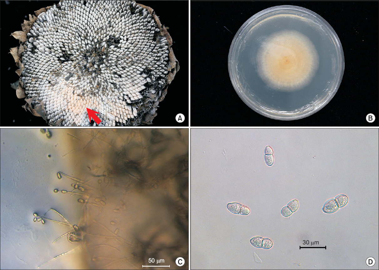
2015ļģä 10ņøö ļåŹņŚģņ£ĀņĀäņ×ÉņøÉņä╝Ēä░ ĒĢ┤ļ░öļØ╝ĻĖ░ ņ×¼ļ░░ĒżņןņŚÉņä£ ĒĢ┤ļ░öļØ╝ĻĖ░ ĻĮā ļČĆļČäņØ┤ Ļ▓ĆĻ▓ī ļ│ĆĒĢśņŚ¼ ĒÖöļó░ ļČĆļČäņØ┤ Ļ░ÉņŚ╝ļÉśĻ│Ā ņżäĻĖ░Ļ╣īņ¦Ć Ļ▓ĆĻ▓ī ļ│ĆĒĢśņśĆļŗż. ņóģņ×ÉņØś Ļ▓ĮņÜ░ļŖö ņä▒ņłÖĒĢśņ¦Ć ļ¬╗ĒĢśņśĆĻ│Ā ņŗ¼ĒĢ£ Ļ▓ĮņÜ░ ņóģņ×ÉĻ░Ć ĻĘĀņé¼ņŚÉ ļŹ«ņśĆņ£╝ļ®░, ļČäĒÖŹņāēņØś Ēżņ×ÉņĖĄņØ┤ ĒśĢņä▒ļÉśĻĖ░ļÅä Ē¢łļŗż(Fig. 1A). ļ│æņØ┤ ņ¦äņĀäļÉ©ņŚÉ ļö░ļØ╝ ņżäĻĖ░Ļ░Ć Ļ▓ĆĻ▓ī ļ│ĆĒĢśņŚ¼ Ļ│Āņé¼ĒĢśņśĆņ£╝ļ®░, ņóģņ×ÉļŖö ņŚ¼ļ¼╝ņ¦Ć ļ¬╗ĒĢśĻ│Ā ņŁēņĀĢņØ┤ļĪ£ ļé©Ļ▒░ļéś ĒāłļØĮĒĢśņśĆļŗż. ņØ┤ļ¤¼ĒĢ£ ņ”ØņāüņØĆ Ēżņן ļé┤ņŚÉņä£ ņŚ¼ļ¤¼ Ļ││ņŚÉ ņé░ļ░£ņĀüņ£╝ļĪ£ ļéśĒāĆļé¼ļŗż.
Fig.┬Ā1
Symptoms of pink rot on sunflower and microscopic investigation of causal organism. (A) Arrow indicates fungal mass in sunflower head. (B) Colony of isolated fungus on potato dextrose agar (PDA) media at 6 days after inoculation at 25┬░C. (C) Conidiophores and spores isolated fungus on PDA media. (D) Spores of isolated fungus on PDA media.
ļ│æņøÉĻĘĀņØś ļČäļ”¼
ĒżņןņŚÉņä£ ļ│æļōĀ ņŗØļ¼╝ņ▓┤ļź╝ ņłśņ¦æĒĢśņŚ¼ ļ│æņøÉĻĘĀņØä ņł£ņłśļČäļ”¼ ļ░░ņ¢æĒĢ£ Ēøä ĻĘĀĒĢÖņĀü ĒŖ╣ņä▒Ļ│╝ ļ│æņøÉņä▒ Ļ▓ĆņĀĢņØä ĒĢśņśĆļŗż. ļ│æņøÉĻĘĀņØä ļČäļ”¼ĒĢśĻĖ░ ņ£äĒĢ┤ Ļ░ÉņŚ╝ļÉ£ ĒÖöļó░ ļČĆļČäņØś ņóģņ×Éļź╝ 70% ņŚÉĒāäņś¼Ļ│╝ 1% NaOCl ņÜ®ņĢĪņŚÉņä£ 30ņ┤łĻ░ä Ēæ£ļ®┤ņé┤ĻĘĀĒĢśĻ│Ā ļ®ĖĻĘĀņłśļĪ£ 2ĒÜī ņäĖņ▓Ö Ēøä ļ®ĖĻĘĀļÉ£ ņŚ¼Ļ│╝ņ¦ĆņŚÉņä£ ļ¼╝ĻĖ░ļź╝ ņĀ£Ļ▒░ Ēøä ļ¼╝ĒĢ£ņ▓£ļ░░ņ¦Ć(water agar)ņŚÉ ņ╣śņāüĒĢśņśĆļŗż. 25┬░C ĒĢŁņś©ĻĖ░ņŚÉņä£ 3ņØ╝Ļ░ä ļ░░ņ¢æĒĢ£ ļÆż ĻĘĀņé¼ņ▓┤ ņäĀļŗ©ļČĆļź╝ ļ¢╝ņ¢┤ Ļ░Éņ×ÉĒĢ£ņ▓£ļ░░ņ¦Ć(Difco, Sparks, MD, USA)ņŚÉ ņś«Ļ▓© 25┬░CņŚÉņä£ ļ░░ņ¢æĒĢśņśĆļŗż. ļ░░ņ¦ĆņŚÉņä£ ĒśĢņä▒ļÉ£ Ēżņ×ÉļĪ£ļČĆĒä░ ļŗ©Ēżņ×ÉļČäļ”¼ļź╝ ņłśĒ¢ēĒĢ£ ļÆż ļ░░ņ¢æĒĢśņŚ¼ Ēżņ×Éļź╝ ĒśĢņä▒ņŗ£ņ╝░ļŗż. ĻĘĀņŻ╝ļ│┤Ļ┤ĆĻ│╝ ļ│æņøÉņä▒ Ļ▓ĆņĀĢņØä ņ£äĒĢ┤ ĒśĢņä▒ļÉ£ Ēżņ×Éļź╝ ņłśĻ▒░ĒĢśņŚ¼ 20% glycerolņŚÉ ļäŻĻ│Ā -70┬░C ņ┤łņĀĆņś©ņĀĆņןĻ│ĀņŚÉ ļ│┤Ļ┤ĆĒĢśņśĆņ£╝ļ®░, ļ│æņøÉņä▒ Ļ▓ĆņĀĢ ļ░Å ņŚ╝ĻĖ░ņä£ņŚ┤ ļČäņäØņŚÉ ņé¼ņÜ®ĒĢśņśĆļŗż.
ļ│æņøÉĻĘĀņØś ĻĘĀĒĢÖņĀü ĒŖ╣ņä▒
ļČäļ”¼ļÉ£ ļ│æņøÉĻĘĀņØĆ Ļ░Éņ×ÉĒĢ£ņ▓£ļ░░ņ¦ĆņŚÉņä£ ĒØ░ņāēņØś ĻĘĀņ┤ØņØä ĒśĢņä▒ĒĢśļ®░ ņ×Éļ×Éņ£╝ļ®░, ņØ┤ĒøäņŚÉļŖö ņŻ╝ĒÖ®ņāēņØś ņøÉĒśĢ ņĮ£ļĪ£ļŗłļź╝ ĒśĢņä▒ĒĢśņśĆņ£╝ļ®░ ļÅÖņØ╝ĒĢ£ ņāēĻ╣öņØś ļČäņāØĒżņ×ÉļŹ®ņ¢┤ļ”¼ļź╝ Ļ┤Ćņ░░ĒĢĀ ņłś ņ׳ņŚłļŗż(Fig. 1B). ConidiophoresļŖö 92.9 ╬╝m (62.5-123.1 ╬╝m)ļĪ£ ļŗ©ņł£ĒśĢ ļśÉļŖö ļČäņ¦ĆĒśĢņØ┤ņŚłļŗż(Fig. 1C). ļČäņāØĒżņ×ÉļŖö ļ¼┤ņāēņØś Ļ▓®ļ¦ēņØ┤ ņŚåĻ▒░ļéś ĒĢśļéś ņ׳ļŖö ļæźĻĘ╝ĒāĆņøÉĒśĢ Ēś╣ņØĆ ņä£ņ¢æļ░░ ļ¬©ņ¢æņ£╝ļĪ£ Ēü¼ĻĖ░ļŖö 10.2-21.4-7.5-12.6 ╬╝m (ĒÅēĻĘĀ 16.3-9.7 Ēāå)ņśĆļŗż(Fig. 1D). ĒĢ┤ļ░öļØ╝ĻĖ░ņŚÉņä£ ļČäļ”¼ĒĢ£ ļ│æņøÉĻĘĀ S068ņØĆ ĻĖ░ņĪ┤ņŚÉ ņĢīļĀżņ¦ä Trichothecium roseumĻ│╝ ĒśĢĒā£ņĀüņ£╝ļĪ£ ņ£Āņé¼ĒĢśņśĆļŗż(Table 1) (Oh ļō▒, 2014).
Table┬Ā1
Morphological characteristics of Trichothecium roseum isolated from sunflower and other hosts
| Structure | Character | S068 | T. roseum* |
|---|---|---|---|
| Colony | Color | Pale roseae | Pale roseae |
| ConidiophoresŌĆā | Shape | Simple or branched belowŌĆā | Simple or branched below |
| Length (╬╝m) | 62.5-123.1 | 150-260 | |
| Conidia | Shape | Ellipsoidal, 2 cell | Ellipsoidal, 2 cell |
| Diameter (╬╝m)ŌĆā | 10.2-21.4├Ś7.5-12.6 | 18-22├Ś8-10 |
* Described by Oh et al. (2014).
ļ│æņøÉņä▒ Ļ▓ĆņĀĢ
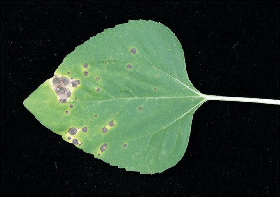
ļ│æņøÉņä▒ Ļ▓ĆņĀĢņØĆ Ēīīņóģ Ēøä 3ņŻ╝ļÉ£ ņ£Āļ¼śņŚÉņä£ ņŗżņŗ£ĒĢśņśĆļŗż. ļČäļ”¼ļÉ£ Ļ│░ĒīĪņØ┤ĻĘĀņØä Ļ░Éņ×ÉĒĢ£ņ▓£ļ░░ņ¦ĆņŚÉņä£ 7ņØ╝Ļ░ä ļ░░ņ¢æĒĢśņŚ¼ ļČäņāØĒżņ×Éļź╝ 1├Ś106 conidia/ml ļåŹļÅäļĪ£ ļ¦×ņČöņ¢┤ Ļ▒┤ņĀäĒĢ£ ĒĢ┤ļ░öļØ╝ĻĖ░ ņ£Āļ¼śņŚÉ 10 mlņö® ļČäļ¼┤ĻĖ░ļĪ£ ņé┤ĒżĒĢśņśĆņ£╝ļ®░, ĒĢśļŻ© ļÅÖņĢł Ļ│ĀņŖĄļÅäņāØņןņāüņŚÉņä£ ņĄ£Ļ│Ā ņŖĄļÅäļź╝ ņ£Āņ¦ĆĒĢ£ ļÆż ĻĘĖ Ēøä ņś©ņŗżņŚÉņä£ 20┬░C-25┬░C ņś©ļÅäļź╝ ņ£Āņ¦ĆĒĢśņśĆļŗż. ņĀæņóģ 9ņØ╝ Ēøä ņ×ÄņŚÉņä£ Ļ▓ĆņØĆņāēņØś ņ£żļ¼ĖĒśĢ ļ░śņĀÉņØ┤ ļéśĒāĆļé¼ņ£╝ļ®░(Fig. 2), ņĀæņóģĒĢ£ ĻĘĀņŻ╝ņÖĆ ļÅÖņØ╝ĒĢ£ ĻĘĀņØ┤ ņ×¼ļČäļ”¼ļÉśņŚłļŗż. ļö░ļØ╝ņä£ ļČäļ”¼ĒĢ£ ļ│æņøÉĻĘĀņØĆ ņøÉņØĖļ│æņøÉĻĘĀņ£╝ļĪ£ ĒīÉļŗ©ļÉśņŚłņ£╝ļ®░ ļīĆņĪ░ĻĄ¼ņŚÉņä£ļŖö ļ│æņ¦ĢņØ┤ ļéśĒāĆļéśņ¦Ć ņĢŖņĢśļŗż.
ņŚ╝ĻĖ░ņä£ņŚ┤ ļČäņäØ
ĒśĢĒā£ņĀüņØĖ ĒŖ╣ņä▒ņØä ļÆĘļ░øņ╣©ĒĢśĻĖ░ ņ£äĒĢ┤ ņŚ╝ĻĖ░ņä£ņŚ┤ ļČäņäØņØä ņŗżņŗ£ĒĢśņśĆļŗż. ļČäļ”¼ĒĢ£ ļ│æņøÉĻĘĀņØä Ļ░Éņ×ÉĒĢ£ņ▓£ļ░░ņ¦ĆņŚÉņä£ 1ņŻ╝ņØ╝Ļ░ä ļ░░ņ¢æ Ēøä ribosomal DNA (rDNA)ņØś internal transcribed spacer (ITS) ņśüņŚŁņØś ņŚ╝ĻĖ░ņä£ņŚ┤ņØä ļČäņäØĒĢśņśĆļŗż(White ļō▒, 1990). Genomic DNAļŖö DNeasy Plant Mini Kit (Qiagen, Hilden, Germany)ļź╝ ņØ┤ņÜ®ĒĢśņŚ¼ ļČäļ”¼ĒĢśņśĆņ£╝ļ®░, ITS1/ITS4 primerļź╝ ņØ┤ņÜ®ĒĢśņŚ¼ PCR ņ”ØĒÅŁĒĢśņśĆļŗż(White ļō▒, 1990). ņ”ØĒÅŁ ņé░ļ¼╝ņØĆ ņŚ╝ĻĖ░ņä£ņŚ┤ ļČäņäØņØä ĒĢśņŚ¼ GenBank database (http://blast.ncbi.nlm.nih.gov/Blast.cgi)ņØś ļŹ░ņØ┤Ēä░ļ▓ĀņØ┤ņŖżļź╝ ņØ┤ņÜ®ĒĢśņŚ¼ ĒÖĢņØĖĒĢśņśĆļŗż. Ļ│äĒåĄņłś ļČäņäØņØĆ GenBank ļŹ░ņØ┤Ēä░ļ▓ĀņØ┤ņŖżņØś ITS ņŚ╝ĻĖ░ņä£ņŚ┤ļōżņØä ņØ┤ņÜ®ĒĢśņŚ¼ MEGA 6.0 ĒöäļĪ£ĻĘĖļשņØä ĒåĄĒĢ┤ neighbor-joining ļ░®ļ▓Ģņ£╝ļĪ£ phylogenetic ļČäņäØņØä ņłśĒ¢ēĒĢśņśĆļŗż. Sequence distanceļŖö Tajima-Nei parameter modelļĪ£ Ļ│äņé░ĒĢśņśĆļŗż(SaitouņÖĆ Nei, 1987; Tamura ļō▒, 2013). ĒĢ┤ļ░öļØ╝ĻĖ░ņŚÉņä£ ļČäļ”¼ĒĢ£ ļ│æņøÉĻĘĀ S068ņØś ITS ņŚ╝ĻĖ░ņä£ņŚ┤ņØś Ēü¼ĻĖ░ļŖö 582 bpļĪ£ GenBankņŚÉ ĻĖ░ĒāüĒĢśņśĆļŗż(accession no. KX768876). National Center for Biotechnology InformationņØś BLAST search Ļ▓░Ļ│╝ Trichothecium roseumļĪ£ ļō▒ļĪØļÉ£ GenBank accession nos. KP317992, KF897865, JX997437 ļō▒Ļ│╝ 100% ņØ╝ņ╣śĒĢśņśĆļŗż. Ļ│äĒåĄņłśņ×æņä▒ Ļ▓░Ļ│╝ KX768876ņØś ITS ņŚ╝ĻĖ░ņä£ņŚ┤ņØ┤ T. roseumĻ│╝ Ļ░ÖņØĆ Ļ│äĒåĄĻĄ░ņŚÉ ņåŹĒĢ©ņØä ĒÖĢņØĖĒĢĀ ņłś ņ׳ņŚłļŗż(Fig. 3).
Fig.┬Ā3
Phylogenetic analysis of sequence of the internal transcribed spacer ribosomal DNA (rDNA) region of the Trichothecium roseum with closely related strains retrieved from GenBank. The tree was constructed based on the neighbor-joining method with 1,000 replicates. The numbers above the branches represent the bootstrap value. The fungus identified in this study is boldfaced.

T. roseumņŚÉ ņØśĒĢ£ ĒĢ┤ļ░öļØ╝ĻĖ░ ņłśĻ│╝ņŚÉņä£ņØś ļ│æņØĆ ņĪ░ņ¦ĆņĢä, ĒÅ┤ļ×Ćļō£ņŚÉņä£ ļ│┤Ļ│ĀļÉśņ¢┤ ņ׳ņ£╝ļéś(FarrĻ│╝ Rossman, 2016) ņÜ░ļ”¼ļéśļØ╝ņŚÉļŖö ņĢäņ¦ü ļ│┤Ļ│ĀļÉśņ¦Ć ņĢŖņĢśļŗż(The Korean Society of Plant Pathology, 2009). ļö░ļØ╝ņä£ ĒĢ┤ļ░öļØ╝ĻĖ░ ĒÖöļó░ņŚÉņä£ ņŹ®ņØīļ│æņØä ņØ╝ņ£╝ĒéżļŖö ĻĘĀņØä ļČäļ”¼ĒĢśņŚ¼ ĻĘĀĒĢÖņĀü ĒŖ╣ņä▒, ļ│æņøÉņä▒ Ļ▓ĆņĀĢ, ITS rDNA ņŚ╝ĻĖ░ņä£ņŚ┤ ļ╣äĻĄÉļČäņäØ ļō▒ņØś Ļ▓░Ļ│╝ļź╝ ļ░öĒāĢņ£╝ļĪ£ T. roseumņŚÉ ņØśĒĢ£ ĒĢ┤ļ░öļØ╝ĻĖ░ ļČäĒÖŹļ╣øņŹ®ņØīļ│æņØ┤ļØ╝ ļ¬ģļ¬ģĒĢśĻ│Āņ×É ĒĢ£ļŗż.
ņÜöņĢĮ
ĒĢ┤ļ░öļØ╝ĻĖ░ ņ×¼ļ░░ĒżņןņŚÉņä£ ĒÖöļó░ ļČĆļČäņØ┤ ņŹ®ļŖö ņ”ØņāüņØ┤ ļéśĒāĆļé¼ļŗż. ļ│æņ¦ĢņØĆ Ļ░ÉņŚ╝ļÉ£ ĒÖöļó░ ļČĆļČäņØ┤ Ļ░łļ│ĆļÉśņ¢┤ ņżäĻĖ░ļĪ£ ļ▓łņĀĖĻ░öļŗż. Ļ░ÉņŚ╝ļÉ£ ĒÖöļó░ņØś ņóģņ×ÉņŚÉļŖö ņŻ╝ĒÖ®ņāēņØś Ēżņ×ÉļŹ®ņ¢┤ļ”¼ļź╝ Ļ┤Ćņ░░ĒĢĀ ņłś ņ׳ņŚłļŗż. ļ│æņ¦Ģņ£╝ļĪ£ļČĆĒä░ Ļ│░ĒīĪņØ┤ļź╝ ņł£ņłś ļČäļ”¼ĒĢśņŚ¼ Ļ░Éņ×ÉĒĢ£ņ▓£ļ░░ņ¦ĆņŚÉ ļ░░ņ¢æĒĢ£ Ļ▓░Ļ│╝, conidiophoresņŚÉ Ēżņ×Éļź╝ ĒśĢņä▒ĒĢśņŚ¼ ĒØ░ņāēņŚÉņä£ ļČäĒÖŹļ╣øņØä ļØĀņŚłļŗż. ConidiophoresļŖö ļŗ©ņł£ĒśĢ ļśÉļŖö ļČäņ¦ĆĒśĢņ£╝ļĪ£ ĻĖĖņØ┤ļŖö 62.5-123.1 ĒāåņśĆļŗż. ļČäņāØĒżņ×ÉļŖö ļ¼┤ņāēņØś Ļ▓®ļ¦ēņØ┤ ņŚåĻ▒░ļéś ĒĢśļéś ņ׳ļŖö ļæźĻĘ╝ĒāĆņøÉĒśĢņŚÉņä£ ņä£ņ¢æļ░░ ļ¬©ņ¢æņ£╝ļĪ£ Ēü¼ĻĖ░ļŖö 10.2-21.4-7.5-12.6 ĒāåņśĆļŗż. ņØ┤ ĻĘĀņØĆ Ļ▒┤ņĀäĒĢ£ ĒĢ┤ļ░öļØ╝ĻĖ░ ņ×ÄņŚÉ ņĀæņóģĒĢśņśĆņØä ļĢī ņ×ÄņŚÉņä£ ņ£żļ¼ĖĒśĢņØś Ļ▓ĆņØĆņāē ļ░śņĀÉņØä ĒśĢņä▒ĒĢśņśĆļŗż. ĻĘĀĒĢÖņĀü ĒŖ╣ņ¦Ģ, ļ│æņøÉņä▒ Ļ▓ĆņĀĢ, ITS ņŚ╝ĻĖ░ņä£ņŚ┤ ļČäņäØ ļō▒ņØś Ļ▓░Ļ│╝ļź╝ ļ░öĒāĢņ£╝ļĪ£ Trichothecium roseumņ£╝ļĪ£ ļÅÖņĀĢļÉśņŚłņ£╝ļ®░ ĒĢ┤ļ░öļØ╝ĻĖ░ ļČäĒÖŹļ╣øņŹ®ņØīļ│æņ£╝ļĪ£ ļ¬ģļ¬ģĒĢśĻ│Āņ×É ĒĢ£ļŗż.
 PDF Links
 PubReader
 Full text via DOI
 Download Citation
 Print
Print


